Diacyclops hanguk
| Notice: | This page is derived from the original publication listed below, whose author(s) should always be credited. Further contributors may edit and improve the content of this page and, consequently, need to be credited as well (see page history). Any assessment of factual correctness requires a careful review of the original article as well as of subsequent contributions.
If you are uncertain whether your planned contribution is correct or not, we suggest that you use the associated discussion page instead of editing the page directly. This page should be cited as follows (rationale):
Citation formats to copy and paste
BibTeX: @article{Karanovic2013ZooKeys267, RIS/ Endnote: TY - JOUR Wikipedia/ Citizendium: <ref name="Karanovic2013ZooKeys267">{{Citation See also the citation download page at the journal. |
Ordo: Cyclopoida
Familia: Cyclopidae
Genus: Diacyclops
Name
Diacyclops hanguk Karanovic & Grygier & Lee, 2013 sp. n. – Wikispecies link – ZooBank link – Pensoft Profile
Type locality
South Korea, Gangwondo, Pyeongchang city, Jinbu, Namhan River, 37°36'56.9"N, 128°32'23.2"E, interstitial water from sandy banks.
Type material
Holotype female dissected on two slides (NIBRIV0000232661). Allotype male from type locality dissected on one slide (NIBRIV0000232662). Other paratypes from type locality: three males and three females on one SEM stub (NIBRIV0000232648), one female dissected on one slide (NIBRIV0000232663), and one male dissected on one slide (NIBRIV0000232664); all collected 12 June 2010, leg. J.-L. Cho.
Additional paratypes: one male and six females together in alcohol (NIBRIV0000232665), from South Korea, Gangwondo, Wonju city, Buron River, 37°14'01.23"N, 127°44'58.78"E, interstitial water from sandy banks, 24 June 2010, leg. J.-L. Cho.
Additional paratypes: five males, two females, and one copepodid together on one SEM stub (NIBRIV0000232653), from South Korea, Gangwondo, Yeongwol city, Namhan River, 37°06'56.9”N, 128°32'23.2”E, interstitial water from sandy banks, 12 June 2010, leg. J.-L. Cho.
Additional paratypes: one male and six females together in alcohol (NIBRIV0000232667), from South Korea, Jeollanamdo, Gurye city, Seomjin River, 35°11'25.4"N, 127°23'00.7"E, interstitial water from sandy banks, 19 June 2010, leg. J.-L. Cho.
Additional paratypes: one male, two females and one copepodid together in alcohol (NIBRIV0000232668), from South Korea, Gangwondo, Wonju city, Jijeong, Seom River, 37°23'10.16"N, 127°51'08.39"E, interstitial water from sandy banks, 24 June 2010, leg. J.-L. Cho.
Additional paratypes: two females and one copepodid together on one SEM stub (NIBRIV0000232653), from South Korea, Jeollanamdo, Gurye city, Yangcheon, Seomjin River, 35°12'04.7"N, 127°35'29.3"E, interstitial water from sandy banks, 19 June 2010, leg. J.-L. Cho.
Etymology
The species name is a phonetic approximation in Latin letters of the country name “Korea” in the Korean language, to be treated as a Latin noun in apposition to the generic name.
Description
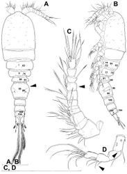
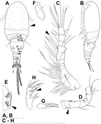
Female (based on holotype and four paratypes from type locality). Total body length, measured from tip of rostrum to posterior margin of caudal rami (excluding caudal setae), from 412 to 445 µm (440 µm in holotype). Preserved specimens colourless; no live specimens observed. Integument relatively weakly sclerotised, smooth, without cuticular pits or cuticular windows. Surface ornamentation of somites consisting of 44 pairs of sensilla and pores and four unpaired (mid-dorsal) pores (those pores and sensilla probably homologous with those of Diacyclops ishidai indicated with same Arabic numerals; those homologous with those of Diacyclops parasuoensis indicated with Roman numerals; presumably novel pores and sensilla indicated with Greek letters consecutively from anterior to posterior end of body, and from dorsal to ventral side in Figs 18A, B, 19A, B, 20A, B, C); no spinules except on anal somite, caudal rami, and appendages. Habitus (Fig. 18A, B) relatively slender, only slightly dorso-ventrally compressed, with prosome/urosome length ratio 1.4 and greatest width in dorsal view at first third of cephalothorax, body prominently arched backwards between prosome and urosome. Body length/width ratio about 3.3 (dorsal view); cephalothorax 1.84 times as wide as genital double-somite. Free pedigerous somites without lateral or dorsal expansions, all connected by well developed arthrodial membranes and having narrow and smooth hyaline fringes. Pleural areas of cephalothorax and free pedigerous somites very short, not covering insertions of cephalic appendages or praecoxae of swimming legs in lateral view.
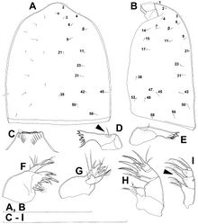
Rostrum (Fig. 19A, B) well developed, membranous, not demarcated at base, broadly rounded and furnished with one pair of frontal sensilla (no. 1).
Cephalothorax (Figs 18A, B, 19A, B) relatively small, 1.2 times as long as its greatest width (dorsal view), widest at posterior third and gently tapering anteriorly and posteriorly, only slightly oval; representing 35% of total body length (together with rostrum). Surface of cephalic shield ornamented with one unpaired dorsal pore (α) and 25 pairs of long sensilla (nos. 2-4, 6, β, 7, 9, 11, 14, 15, 17, 21, 23, 31, 38, 39, 42, 45, 47, 48, 50, 52, 56, 58); sensilla pair no. 39 highly asymmetrical; sensilla and pores 39-58 belonging to first pedigerous somite, latter being incorporated into cephalothorax.
Second pedigerous somite (Figs 18A, B) well developed, only slightly narrower than cephalothorax and tapering posteriorly, unornamented.
Third pedigerous somite (Fig. 18A, B) shorter and narrower than second in dorsal view, widest at midlength in dorsal view and with slightly flared latero-posterior corners, ornamented with one unpaired dorsal pore (no. I) and four pairs of large sensilla (nos. 63, 64, 72, 74).
Fourth pedigerous somite (Fig. 18A, B) significantly shorter and narrower than third, with slightly flared latero-posterior corners, nicely rounded in dorsal view, ornamented with only one unpaired dorsal pore (no. II) and two pairs of large sensilla (nos. 75, 77); recognition of serially homologous pairs relatively easy (probably II=I, 75=63, 77=72).
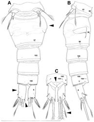
Fifth pedigerous somite (Figs 18A, B, 20A, B) short, significantly narrower than fourth pedigerous somite or genital double-somite in dorsal view, with prominently flared latero-posterior corners, ornamented with two pairs of large dorsal sensilla (nos. 80, 81); recognition of serially homologous pairs relatively easy, i.e. 80=75 and 81=77; hyaline fringe very narrow and smooth.
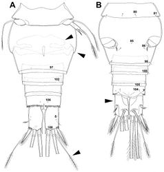
Genital double-somite (Figs 18A, 20A, B) large but proportionately short (arrowed in Fig. 20A), with deep lateral recesses at level of sixth legs and only slightly swollen antero-ventrally, widest at first third of its length in dorsal (or ventral) view and gradually tapering posteriorly, 0.7 times as long as its greatest width (dorsal view); ornamented with one unpaired dorsal central pore (no. 85), one pair of central dorsal sensilla (no. 86), one pair of posterior lateral sensilla (no. 96), and one pair of ventral posterior pores (no. 97); central dorsal sensilla probably serially homologous to those on fifth pedigerous somite (i.e. 86=80), but posterior sensilla and pores without homologous pairs. Copulatory pore small, oval, situated at about 3/5 of genital double-somite ventrally; copulatory duct relatively wide, siphon-shaped and directed anteriorly, weakly sclerotised. Hyaline fringe wavy, not serrated. Seminal receptacle anvil-shaped, with relatively short anterior expansion and long lateral arms, constricted at middle, and with equally long and wide posterior expansion, together representing 52% of double-somite’s length; ovipores situated dorso-laterally at midlength of double-somite, covered by reduced sixth legs.
Third urosomite (Figs 18A, 20A, B) relatively short, about 1.9 times as wide as long and 0.4 times as long as genital double-somite in dorsal view, also with wavy hyaline fringe, ornamented with one pair of lateral posterior sensilla (no. 100) and one pair of ventral posterior pores (no. 102); serially homologous pores and sensilla easy to recognize, i.e. 100=96 and 102=97.
Preanal urosomite (Figs 18A, 20A, B) slightly narrower and shorter than third, also with wavy hyaline fringe, unornamented.
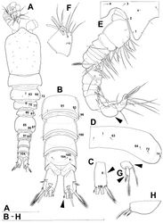
Anal somite (Figs 18A, 20A, B, C) slightly narrower and significantly shorter than preanal, with short medial cleft, ornamented with one pair of large dorsal sensilla (no. 104), two pairs of dorso-lateral pores (nos. 105, γ), one pair of small ventral pores (no. 106), and continous posterior row of large spinules. Anal sinus with two diagonal rows of short, slender spinules. Anal operculum very wide (arrowed in Fig. 20C), slightly convex, reaching posterior margin of anal somite, and representing 54% of anal somite’s width.
Caudal rami (Fig. 20A, B, C) very short (arrowed in Fig. 20A), almost cylindrical and parallel, inserted very close to each other (space between them less than half of width of ramus), with deep dorso-median anterior depression (as continuation of anal sinus), and with narrower base than rest of ramus (particularly in ventral view); rami approximately 1.8 times as long as wide (ventral view) and 1.6 times as long as anal somite, each armed with six setae (one dorsal, one lateral, and four terminal); ornamented with one ventral pore at 1/3 length, one pore on tip of small protuberance on distal ventral margin between two principal terminal setae (no. 108), and rows of small spinules at base of lateral setae. Dorsal seta slender and long (arrowed in Fig. 20C), about 1.4 times as long as ramus, inserted at 5/6 of ramus length, biarticulate at base (inserted on small pseudo-joint) and pinnate distally. Lateral seta small, inserted dorso-laterally at 2/3 of ramus length, about 0.6 times as long as ramus width, unipinnate laterally and uniarticulate at base. Outermost terminal seta stout, spiniform, 0.8 times as long as ramus, densely bipinnate. Innermost terminal seta minute (arrowed in Fig. 20A), sparsely pinnate, 0.2 times as long as outermost terminal seta. Two principal terminal setae with breaking planes, bipinnate; inner one about 1.5 times as long as outer one and 5.3 times as long as caudal rami.
Antennula (Fig. 18C) 11-segmented, with very short eighth segment (arrowed in Fig. 18C), slightly curved along caudal margin, directed laterally, not reaching posterior margin of cephalothoracic shield, ornamented only with proximo-ventral arc of spinules on first segment (no pits or other integumental structures), with armature formula as in Diacyclops ishidai. Only one seta on tenth segment with breaking plane, no seta biarticulate basally, and most larger setae sparsely pinnate distally; both aesthetascs very slender, that on eighth segment reaching posterior margin of ninth segment. One seta on fourth and one on fifth segment spiniform and short; all other setae slender; one apical seta on eleventh segment fused basally to aesthetasc. Length ratio of antennular segments, from proximal end and along caudal margin, 1 : 0.4 : 0.6 : 0.3 : 0.2 : 0.5 : 0.9 : 0.7 : 0.5 : 0.7 : 1.
Antenna (Figs 18D, 21E) five-segmented, strongly curved along caudal margin, comprising extremely short coxa, much longer basis, and three-segmented endopod. Coxa without armature or ornamentation, about 0.2 times as long as wide. Basis cylindrical, 1.5 times as long as wide, ornamented with three short rows of three or four spinules each on ventral surface, armed with only one seta on distal inner corner (exopodal seta absent). First endopodal segment slightly narrowed at base and with small expansions on caudal margin but generally cylindrical, 1.4 times as long as wide and 0.9 times as long as basis, with smooth inner seta at 2/3 length and row of minute spinules caudo-dorsally. Second endopodal segment also with narrowed basal part, 1.6 times as long as wide, about as long as first endopodal segment, bearing only five smooth setae along inner margin (arrowed in Fig. 18D), ornamented with one row of spinules along caudal margin. Third endopodal segment cylindrical, 1.9 times as long as wide and slightly shorter than second endopodal segment, ornamented with two rows of slender spinules along caudal margin and armed with seven smooth apical setae (four of them strong and geniculate).
Labrum (Fig. 19C) a relatively large trapezoidal plate, with muscular base and strongly sclerotised distal margin (cutting edge), ornamented with two diagonal rows of seven long and slender spinules each on anterior surface. Cutting edge almost straight, with 13 more or less sharp teeth between produced and rounded lateral corners.
Mandibula (Fig. 19D, E) composed of coxa and minute palp. Cutting edge of coxal gnathobase with four slender spinules on anterior surface, five apical teeth, and dorsalmost unipinnate seta. Ventralmost tooth strongest and quadricuspidate, second and third teeth from ventral side bicuspidate, two dorsalmost teeth unicuspidate and partly fused basally. Palp represented by extremely small but distinct segment, unornamented, armed with single short and smooth apical seta (arrowed in Fig. 19D).
Maxillula (Fig. 19F, G) composed of praecoxa and one-segmented large palp, unornamented. Praecoxal arthrite bearing four very strong and smooth distal spines (three blunt and fused at base, one distinct at base and sharp) and six medial elements (proximalmost one longest and plumose, two distalmost ones large and strong, three in between small and slender). Palp composed of coxobasis and one-segmented endopod, with latter fused basally to coxobasis. Coxobasis with short proximal seta (probably representing exopod) and three medial setae (two slender and smooth, one strong and pinnate). Endopod with three slender, smooth setae.
Maxilla (Fig. 19H) 5-segmented but praecoxa partly fused to coxa on anterior surface, unornamented. Proximal endite of praecoxa robust, armed with two sparsely bipinnate setae; distal endite slightly smaller than proximal one and unarmed. Proximal endite of coxa with one bipinnate seta; distal endite highly mobile, elongated and armed apically with two setae, proximal one considerably longer and stronger than distal one. Basis expanded into robust and smooth claw, armed with two setae; strong seta densely pinnate along convex (ventral) margin, robust and spiniform; small seta smooth and slender, inserted on posterior surface. Endopod two-segmented but segmentation not easily discernable; proximal segment armed with two robust, unipinnate setae; distal segment with one robust, unipinnate apical seta and two slender subapical setae. Longest seta on distal endopodal segment 0.8 times as long as longer seta on proximal segment. All strong setae on basis and endopod, as well as basal claw, unguiculate.
Maxilliped (Fig. 19I) four-segmented, composed of syncoxa, basis, and two-segmented endopod; second endopodal segment minute; basis with only one armature element (arrowed in Fig. 19I). Ornamentation consisting of two rows of long, slender spinules on basis (one row on posterior surface, other on anterior surface), as well as four spinules on anterior surface of first endopodal segment. Armature formula: 2.1.1.2. All inner setae pinnate, very strong, and unguiculate.
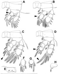
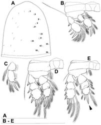
All swimming legs (Fig. 21A, B, C, D, F, G) relatively small, with segmentation formula as in Diacyclops parasuoensis, as well as spine and setal formulae of ultimate exopodal segment, but with some differences in armature of endopods, ornamentation of different segments, and proportions of some segment and armature elements. Exopods slightly longer than endopods on all legs. All setae on endopods and exopods slender and plumose, except apical seta on exopod of first leg pinnate along outer margin and plumose along inner (Fig. 21A); no modified setae observed. All spines strong and bipinnate. Intercoxal sclerite of all swimming legs with slightly concave distal margin and lacking any surface ornamentation.
First swimming leg (Fig. 21A) shorter than other swimming legs; praecoxa unarmed, ornamented with distal row of minute spinules on anterior surface; coxa twice as wide as long, ornamented with distal row of large spinules on anterior surface and small pore on anterior surface close to inner margin, armed with slender plumose seta on inner-distal corner; basis almost pentagonal, 0.7 times as long as coxa, armed with long and slender seta outer seta and strong and short inner-distal element (latter not reaching distal margin of first endopodal segment, i.e. much shorter than in Diacyclops ishidai or Diacyclops parasuoensis;arrowed in Fig. 21A); inner margin smooth, ornamented with two posterior rows of minute spinules on anterior surface (one at base of inner seta, other at base of endopod), and one cuticular pore on anterior surface close to outer margin; exopod and endopod armed as in Diacyclops parasuoensis, but second segments more elongated, and second endopodal segment with inner notch and shorter middle inner seta.
Second swimming leg (Fig. 21B, F) similar to that of Diacyclops parasuoensis, but second endopodal segment with only three inner setae (arrowed in Fig. 21B), with or without inner notch (showing original segmentation), and third exopodal segment with all setae proportionatelly much shorter than in Diacyclops parasuoensis; apical spine on second endopodal segment 0.8 times as long as segment and 0.7 times as long as inner distal seta; second endopodal segment about 1.4 times as long as wide and 1.3 times as long as first endopodal segment.
Third swimming leg (Fig. 21C) similar to that of Diacyclops parasuoensis, but coxa without ornamentation on posterior surface, endopodal setae proportionately shorter (arrowed in Fig. 21C), and setae on third exopodal segment more obviously progressively longer from distal to proximal; apical spine on third endopodal segment 1.1 times as long as segment and 0.7 times as long as apical seta; third endopodal segment about as long as wide and 1.3 times as long as second endopodal segment.
Fourth swimming leg (Fig. 21D, G) generally similar to that of Diacyclops parasuoensis, but intercoxal sclerite unornamented, coxa without proximal row of spinules on posterior surface, proximal seta on third endopodal segment much shorter (arrowed in Fig. 21D), and proximal seta on third exopodal segment much longer (arrowed in Fig. 21D); third endopodal segment only about 0.9 times as long as wide, and only as long as second endopodal segment; inner apical spine on third endopodal segment 1.4 times as long as outer apical spine, about as long as segment, and less than half as long as distal inner seta; apical spines parallel.
Fifth leg (Fig. 20A, B) inserted ventrally, relatively small, two-segmented, with same armature as in previous four species, but with very different shape and proportions. Protopod relatively wide (although not as wide as in Diacyclops ishidai), almost rhomboidal, about 0.65 times as long as greatest width, unornamented, armed with single distally unipinnate and slender outer seta inserted on short setophore. Exopod much narrower than protopod, almost cylindrical, 1.1 times as long as protopod and twice as long as wide, unornamented, armed with long apical seta and subapical inner spine; apical seta bipinnate distally, as long as basal seta, 2.8 times as long as exopod, and 2.7 times as long as subapical spine, reaching midlength of genital double-somite; subapical exopodal spine strong, bipinnate, 0.9 times as long as exopod and almost twice as long as exopod’s greatest width.
Sixth leg (Fig. 21H) small semicircular cuticular plate, unornamented, armed with two short, smooth spines and, external to these, one longer and distally bipinnate seta; inner spine fused to plate, outer one articulated basally; outermost seta directed postero-dorsally.
Male (based on allotype and five paratypes from type locality). Total body length from 380 to 405 µm (383 µm in allotype). Urosome with free genital somite. Habitus (Fig. 22A) more slender than in female, with prosome/urosome length ratio about 1.5 and greatest width in dorsal view at first third of cephalothorax. Body length/width ratio 3.9; cephalothorax about 1.7 times as wide as genital somite. Cephalothorax 1.3 times as long as wide and slightly tapering towards posterior margin in dorsal view; representing 34% of total body length; dorsal sensilla pair no. 39 symmetrical. Ornamentation of cephalothorax (Fig. 22A), free prosomites (Fig. 22D), and first and last three urosomites (Fig. 22A, B) with same number and distribution of sensilla and pores as in female.
Genital somite (Fig. 22A) 1.5 times as wide as long in dorsal view, with bluntly serrated hyaline fringe dorsally, ornamented with one unpaired dorsal pore (no. 85), one pair of dorsal sensilla (nos. 86), and one pair of dorsolateral pores (no. 87; N.B., these absent in female); no spermatophores visible inside. Third urosomite (Fig. 22B) homologous to posterior part of female genital double-somite, ornamented with ventral pair of posterior pores (no. 97) and lateral pair of sensilla (no. 96) as in female, but additionally with dorsal unpaired pore (no. 91) and dorsal pair of sensilla (no. 93).
Caudal rami (Fig. 22B, C) slightly more divergent than in female, but equally short, with minute innermost terminal seta (arrowed in Fig. 22B) and with anterior ventral pore (δ; arrowed in Fig. 22C).
Antennula (Fig. 22E, F) strongly prehensile and digeniculate, 17-segmented (but with sixteenth and seventeenth segments partly fused), ornamented with spinules only on first segment (as in female), with anvil-shaped cuticular ridges on anterior margin of fourteenth and fifteenth segments (distal geniculation), with extremely short sixteenth segment (arrowed in Fig. 22E). Armature formula as follows: 8+3ae.4.2.2+ae.2.2.2.2.2+ae.2.2.2.2 + ae.2.1.4.7+ae. All aesthetascs linguiform, slender and short, apical one on sixteenth segment fused basally to one seta.
Antenna, labrum, mandibula, maxillula, maxilla, swimming legs, and fifth leg as in female.
Sixth leg (Fig. 22H) large cuticular plate, unornamented, armed with small inner spine and two bipinnate setae on outer distal corner; outermost seta 2.2 times as long as inner bipinnate seta, as well as 2.4 times as long as innermost spine.
Remarks
Diacyclops hanguk sp. n. belongs to the languidoides-group based on its 11-segmented antennula and the swimming leg segmentation formula (exopod/endopod) 2/2, 3/2, 3/3, 3/3. Detailed examination of the pattern of pores and sensilla on the somites and the armature of the mouth appendages show that this species is only distantly related to all other East Asian species of this species-group except for Diacyclops parahanguk sp. n. from Japan (see below). It it quite clear that the two form a sibling species pair, with their extremely short caudal rami and genital double somite, extremely wide anal operculum, almost identical pattern of pores and sensilla on all somites, minute innermost terminal caudal seta, long dorsal caudal seta, wider than long eighth segment of the antennula, extremely reduced armature formula of the antenna (1.1.5.7), single seta on the mandibular palp, identical armature formula of the swimming legs (with only three inner setae on the second endopodal segment of the first leg), extremely short third endopodal segment of the fourth leg, and progressively longer setae from the outer side on the third exopodal segment of the fourth leg. The two species can be distinguished by their habitus, armature of the maxillipedal basis, ornamentation of the labrum and antennal basis, relative length of the aesthetasc on the eighth antennular segment, small differences in the shape of the seminal receptacle, genital double-somite, and caudal rami, and relative lengths of the dorsal caudal seta and inner apical spine on the third endopodal segment of the fourth leg (all these arrowed in Figs 23–25). Additionally, small differences were observed in the pattern of pores and sensilla, with sensilla no. 21 less widely spaced in Diacyclops hanguk.
No other species of the languidoides-group, nor any other species of Diacyclops, has such short caudal rami and minute innermost terminal caudal setae. These characters are traditionally only found in much more reduced cyclopoids (usually with variously reduced fifth legs), such as members of the genus Itocyclops Reid & Ishida, 2000 from Japan and North America (see Reid and Ishida 2000[1]). It would be easy to speculate how this monospecific genus could have originated from an acestor similar to Diacyclops hanguk through a series of reductions in both the swimming leg segmentation and the armature and segmentation of the fifth leg, if Itocyclops did not exibit a more plesiomorphic armature of the antenna and maxilliped.
The only other described Diacyclops with similarly short caudal rami is the Romanian Diacyclops languidoides spelaeus Pleşa, 1956. This species does not even belong to the languidoides-group, as is shown by the three-segmented exopod of its first leg (see Pleşa 1956[2]), even though Dussart and Defaye (2006)[3] recently still listed it as a subspecies of Diacyclops languidoides (Lilljeborg, 1901). The latter authors mentioned that Diacyclops languidoides spelaeus could be a junior synonym of Diacyclops stygius deminutus (Chappuis, 1925), following Monchenko (1974)[4], but this is just a speculation until both taxa are properly redescribed. Both differ from Diacyclops hanguk by the segmentation of the swimming legs, and by their much longer innermost terminal caudal seta.
Original Description
- Karanovic, T; Grygier, M; Lee, W; 2013: Endemism of subterranean Diacyclops in Korea and Japan, with descriptions of seven new species of the languidoides-group and redescriptions of D. brevifurcus Ishida, 2006 and D. suoensis Ito, 1954 (Crustacea, Copepoda, Cyclopoida) ZooKeys, 267: 1-76. doi
Other References
- ↑ Reid J, Ishida T (2000) Itocyclops, a new genus proposed for Speocyclops yezoensis (Copepoda: Cyclopoida: Cyclopidae). Journal of Crustacean Biology 20: 589-596. doi: [0589:IANGPF2.0.CO;2 10.1651/0278-0372(2000)020[0589:IANGPF]2.0.CO;2]
- ↑ Pleşa C (1956) Contributii la fauna ciclopidelor (crustacee copepode) din Republica Populara Romina. Analele Institutului de Cercetari Piscicole 1: 363-372.
- ↑ Dussart B, Defaye D (2006) World Directory of Crustacea Copepoda of Inland Waters, II – Cyclopiformes. Backhuys Publishers, Leiden, 352 pp.
- ↑ Monchenko V (1974) Schelepnoroti Ciklopopodibni Ciklopi (Cyclopidae). Fauna Ukraïni 27(3). Kiev, 452 pp.
Images
|
