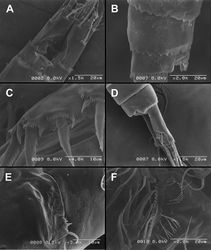
Diacyclops ishidai
| Notice: | This page is derived from the original publication listed below, whose author(s) should always be credited. Further contributors may edit and improve the content of this page and, consequently, need to be credited as well (see page history). Any assessment of factual correctness requires a careful review of the original article as well as of subsequent contributions.
If you are uncertain whether your planned contribution is correct or not, we suggest that you use the associated discussion page instead of editing the page directly. This page should be cited as follows (rationale):
Citation formats to copy and paste
BibTeX: @article{Karanovic2013ZooKeys267, RIS/ Endnote: TY - JOUR Wikipedia/ Citizendium: <ref name="Karanovic2013ZooKeys267">{{Citation See also the citation download page at the journal. |
Ordo: Cyclopoida
Familia: Cyclopidae
Genus: Diacyclops
Name
Diacyclops ishidai Karanovic & Grygier & Lee, 2013 sp. n. – Wikispecies link – ZooBank link – Pensoft Profile
Type locality
Japan, Shiga prefecture, Otsu city, border of Kamitanakami-Nakano-cho township and Shinme 2-chome district, Kisshoji River about 0.7 km upstream from outflow into Daido River, 34°56.732'N, 135°57.331'E, interstitial water from coarse sand and gravel.
Type material
Holotype female dissected on two slides (LBM 1430005377). Allotype male from type locality also dissected on two slides (LBM 1430005378). Other paratypes from type locality: three females on one SEM stub (LBM1430005379), two females dissected on one slide each (LBM 1430005380, LBM 1430005381), and one male and four females together in ethanol (LBM 1430005382), all collected 27 September 2009, leg. T. Karanovic.
Additional paratypes: eight females in ethanol (LBM 1430005383) from Japan, Shiga prefecture, Otsu city, Nakano 3-chome township, Daido River, 34°57.043'N, 135°57.044'E, interstitial water from medium to coarse sand, 27 September 2009, leg. T. Karanovic.
Etymology
The new species is named in honour of the late Dr. Teruo Ishida, in recognition of his contribution to our knowledge of freshwater copepods in Japan. The name is a noun in the genitive singular.
Description
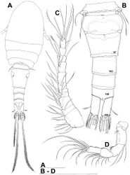
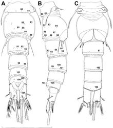
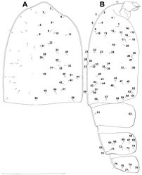
Female (based on holotype and five paratypes from type locality). Total body length, measured from tip of rostrum to posterior margin of caudal rami (excluding caudal setae), from 450 to 482 µm (453 µm in holotype). Preserved specimens colourless; no live specimens observed. Integument relatively weakly sclerotised, smooth, without cuticular pits or cuticular windows. Surface ornamentation of somites consisting of 92 pairs and seven unpaired (mid-dorsal) pores and sensilla (numbered with Arabic numerals consecutively from anterior to posterior end of body, and from dorsal to ventral side in Figs 1B, 2A, B, 3F; but illustrated in more detail for male specimens, see Figs 4, 5, 6); no spinules except on anal somite, caudal rami, and appendages. Habitus (Fig. 1A) relatively robust, not dorso-ventrally compressed, with prosome/urosome length ratio 1.3 and greatest width in dorsal view at posterior end of cephalothorax. Body length/width ratio about 3 (dorsal view); cephalothorax 2.1 times as wide as genital double-somite. Free pedigerous somites without lateral or dorsal expansions, all connected by well developed arthrodial membranes and all with narrow and smooth hyaline fringes. Pleural areas of cephalothorax and free pedigerous somites relatively well developed, covering insertions of cephalic appendages and praecoxae and partly covering coxae of swimming legs in lateral view.
Rostrum well developed, membranous, not demarcated at base, broadly rounded and furnished with single frontal pair of sensilla (no. 1).
Cephalothorax (Fig. 1A) large, 1.1 times as long as its greatest width (dorsal view), narrower at anterior part and perfectly oval; representing 38% of total body length. Surface of cephalic shield ornamented with three unpaired mid-dorsal sensilla and pores (nos. 5, 8, 55) and 56 pairs of long sensilla and small cuticular pores (nos. 2-5, 6, 7, 9-54, 56-60); pores and sensilla 39-60 belonging to first pedigerous somite, latter being incorporated into cephalothorax.
Second pedigerous (first free) somite (Figs 1A) relatively short, tapering posteriorly, ornamented with just one pair of dorsal sensilla (no. 61) and one pair of lateral pores (no. 62); serially homologous pairs impossible to establish.
Third pedigerous somite (Fig. 1A) slightly longer than second and significantly narrower in dorsal view, ornamented with 12 pairs of large sensilla (nos. 63-74); recognition of serially homologous pairs not easy, but probably dorsolateral pair of sensilla no. 64 serially homologous to pair no. 61 on second pedigerous somite.
Fourth pedigerous somite (Figs 1A) significantly shorter and narrower than third, with slightly flared latero-posterior corners and only five pairs of large sensilla (nos. 75–77); recognition of serially homologous pairs slightly easier than for two previous prosomites (probably 75=63, 76=71, 77=72, 78=73, 79=74).
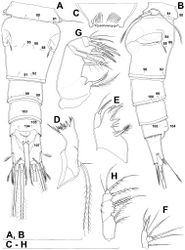
Fifth pedigerous (first urosomal) somite (Figs 1B, 2A, B) short, significantly narrower than fourth pedigerous somite and also narrower than genital double-somite in dorsal view, ornamented with two pairs of large dorsal sensilla (nos. 80, 81); recognition of serially homologous pairs easy, i.e. 80=75 and 81=77; hyaline fringe very narrow, smooth or barely visibly serrated.
Genital double-somite (Figs 1B, 2A, B, 3F) large, swollen antero-ventrally with deep lateral recesses at level of sixth legs, widest at first quarter of its length and gradually tapering posteriorly, only slightly longer than its greatest width (dorsal view), ornamented with one unpaired central dorsal pore (no. 85), two pairs of central dorsal sensilla (nos. 86, 88), one pair of lateral central pores (no. 84), one unpaired posterior dorsal pore (no. 91), one pair of posterior sensilla (no. 92), and two pairs of ventro-lateral posterior pores (nos. 96, 97); central dorsal sensilla probably serially homologous to those on fifth pedigerous somite (i.e. 86=80, 88=81), but recognition of serial homologies of posterior sensilla and pores much harder (perhaps 91=85, 92=86); hyaline fringe deeply and irregularly serrated. Copulatory pore very small, oval, situated ventrally at about midlength of double-somite; copulatory duct narrow, siphon-shaped, weakly sclerotised. Seminal receptacle characteristically shaped, with relatively large anterior expansion, constriction at midlength, and shorter but broader posterior expansion, altogether representing 49% of double-somite’s length. Ovipores situated dorso-laterally at 1/3 length of double-somite, covered by reduced sixth legs.
Third (ancestral fourth) urosomite (Figs 1B, 2A, B) relatively short, about 1.8 times as wide as long and less than 0.4 times as long as genital double-somite in dorsal view, also with deeply and irregularly serrated hyaline fringe, ornamented with unpaired dorsal posterior pore (no. 98), two pairs of dorso-lateral posterior sensilla (nos. 99, 100), and one pair of ventro-lateral posterior pores (nos. 102); serially homologous pores and sensilla not easy to recognize on genital double-somite, except 98=91.
Fourth (preanal) urosomite (Figs 1B, 2A, B, 26A, B) narrower and shorter than third, also with deeply and irregularly serrated hyaline fringe; ornamented only with unpaired dorsal pore (no. 103), serially homologous to pore no. 98 on third urosomite.
Anal somite (Figs 1B, 2A, B, 26A, B) slightly narrower and significantly shorter than preanal somite, with short medial cleft; ornamented with one pair of large dorsal sensilla (no. 104), one pair of small dorsal pores (no. 105), one pair of small ventral pores (no. 106), continous posterior row of small spinules, and two diagonal parallel rows of somewhat larger spinules on both sides of anal sinus. Anal operculum wide, short, slightly convex, not reaching posterior margin of anal somite, representing 59% of anal somite’s width.
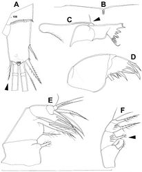
Caudal rami (Figs 1A, B, 2A, B, 26A) cylindrical, parallel, inserted close to each other, with deep dorso-median anterior depression (as continuation of anal sinus), approximately twice as long as wide (ventral view) and twice as long as anal somite; armed with six setae (one dorsal, one lateral, and four terminal); ornamented with one dorsal pore (no. 107), one pore on tip of large protuberance on distal margin ventrally between two terminal setae (no. 108), and rows of small spinules at base of lateral setae. Dorsal seta slender, about as long as ramus, inserted at 5/6 of ramus length, biarticulate at base (inserted on small pseudo-joint), and pinnate distally. Lateral seta inserted at 2/3 of ramus length, 0.4 times as long as dorsal seta, unipinnate laterally and uniarticulate at base. Outermost terminal seta stout, spiniform, 0.7 times as long as ramus, densely bipinnate. Innermost terminal (accessory) seta slightly longer and more slender than outermost terminal seta, sparsely pinnate along outer margin and densely pinnate along inner margin. Principal terminal setae with breaking planes, bipinnate; inner principal terminal seta about 1.8 times as long as outer one and 6.6 times as long as caudal rami.
Antennula (Fig. 1C) 11-segmented, slightly curved along caudal margin, directed postero-laterally, not reaching posterior margin of cephalothoracic shield, ornamented only with proximo-ventral arc of spinules on first segment (no pits or other integumental structures), with armature formula as follows (ae = aesthetasc): 8.4.8.4.2.2.3.2+ae.2.3.7+ae. Only one of terminal seta on ultimate segment biarticulate basally and most of longer setae sparsely pinnate distally; both aesthetascs very slender, that on eighth segment reaching distal margin of ninth segment. One seta on fourth and one on fifth segment spiniform and short, all other setae slender; one apical seta on 11th segment fused basally to aesthetasc. Length ratio of antennular segments, from proximal end and along caudal margin, 1 : 0.3 : 0.9 : 0.4 : 0.3 : 0.6 : 1 : 0.9 : 0.6 : 0.7 : 1.
Antenna (Fig. 1D) five-segmented, strongly curved along caudal margin, comprising very short coxa, much longer basis, and three-segmented endopod. Coxa without armature or ornamentation, about half as long as wide. Basis cylindrical, 1.8 times as long as wide, ornamented with two short diagonal rows of spinules on ventral surface, two transverse rows of small spinules on dorsal surface, and three large spinules along caudal margin, also armed with two subequal smooth setae on distal inner corner (exopodal seta absent). First endopodal segment narrowed basally but generally cylindrical, 1.6 times as long as wide and 0.8 times as long as basis, with smooth inner seta at 2/3 length and patch of large spinules along caudal margin. Second endopodal segment also with narrowed basal part, 1.4 times as long as wide, about 0.8 times as long as first segment, bearing eight smooth setae along inner margin (these progressively longer from proximal to distal), ornamented with one row of spinules along caudal margin. Third endopodal segment cylindrical, 2.3 times as long as wide and slightly longer than second endopodal segment, ornamented with two rows of slender spinules along caudal margin, armed with eight apical setae (four of them strong and geniculate; two pinnate).
Labrum (Fig. 2C) a relatively large trapezoidal plate, with muscular base and strongly sclerotised distal margin (cutting edge), ornamented with two short diagonal rows of nine long and slender spinules each on anterior surface. Cutting edge almost straight, with 11 sharp teeth between produced and rounded lateral corners.
Mandibula (Fig. 2D) composed of coxa and small palp. Cutting edge of coxal gnathobase with five slender spinules on anterior surface, eight apical teeth, and dorsalmost unipinnate seta; ventralmost tooth strongest and quadricuspidate, second and fourth teeth from ventral side bicuspidate, all other teeth unicuspidate; three dorsalmost simple teeth partly fused basally and progressively longer from ventral to dorsal. Palp twice as wide as long, unornamented, armed with three apical setae, two of them long and bipinnate and one short and smooth; pinnate setae subequal in length, directed posteriorly, not reaching posterior margin of cephalic shield.
Maxillula (Fig. 2E, F) composed of praecoxa and two-segmented palp, unornamented. Praecoxal arthrite bearing four very strong distal spines (three of them smooth, blunt, and fused at base; one distinct at base, sharp and with single proximal spinule) and six medial elements (proximalmost one longest and plumose, two most distal ones large and strong, three in between small and slender). Palp composed of coxobasis and one-segmented endopod. Coxobasis with slender proximal seta (probably representing exopod) and three medial setae (two slender, one strong). Endopod with three slender pinnate setae.
Maxilla (Figs 2G) 5-segmented but praecoxa partly fused to coxa on anterior surface, unornamented. Proximal endite of praecoxa robust, armed with two subequal, sparsely bipinnate setae; distal endite small, unarmed. Proximal endite of coxa with one bipinnate seta; distal endite highly mobile, elongated and armed apically with two pinnate setae, proximal one of which considerably longer and stronger. Basis expanded into robust claw; claw furnished with longitudinal row of four spinules at midlength and armed with two setae: strong seta about as long as claw and pinnate, small seta smooth and slender. Endopod two-segmented, but segmentation not easily discernable; proximal segment armed with two robust, unipinnate setae; distal segment with one robust, unipinnate apical seta and two slender and much shorter subapical setae. Longest seta on distal endopodal segment 0.8 times as long as longer seta on proximal endopodal segment. All strong setae on basis and endopod, as well as basal claw, unguiculate.
Maxilliped (Fig. 2H) four-segmented, composed of syncoxa, basis, and two-segmented endopod. Ornamentation consisting of three rows of long, slender spinules on basis (two transverse rows on posterior surface close to outer margin and one longitudinal row on anterior surface close to inner margin), as well as two spinules on anterior surface of first endopodal segment. Armature formula: 2.2.1.3. All inner setae pinnate, very strong, and unguiculate.
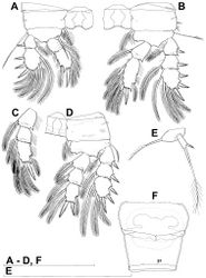
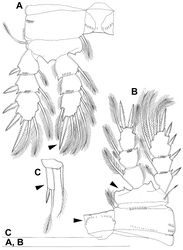
All swimming legs (Figs 3A, B, C, D, 26C) relatively small, composed of minute, triangular praecoxa, large, rectangular coxa, short basis, and slender exopod and endopod. Exopods and endopods approximately equally long in all legs, their segmentation formula (exopod/endopod): 2/2.3/2.3/3.3/3. Ultimate exopodal segment spine formula 3.3.3.3 and setal formula 5.4.4.4. All setae on endopods and exopods slender and plumose, except apical seta on exopod of first leg pinnate along outer margin and plumose along inner (Fig. 3A); no modified setae observed. All spines strong and bipinnate. Intercoxal sclerite of all swimming legs with slightly concave distal margin and lacking surface ornamentation, except on posterior surface of fourth leg.
First swimming leg (Fig. 3A) shorter than other swimming legs; praecoxa unarmed, ornamented with distal row of small spinules on anterior surface; coxa 2.3 times as wide as long, ornamented with short, transverse row of spinules on posterior surface close to outer margin, distal row of minute spinules on anterior surface, and small pore on anterior surface close to inner margin, armed with long, plumose seta on inner-distal corner; basis almost pentagonal, 0.8 times as long as coxa, armed with long, slender outer seta and strong, bipinnate inner-distal element (latter reaching to 2/3 length of second endopodal segment), ornamented with row of slender spinules along inner margin, two posterior rows of shorter and stronger spinules on anterior surface (one at base of inner seta, other at base of endopod), and one cuticular pore on anterior surface close to outer margin; exopod with single outer spine and single inner seta on first segment, with three outer spines and five setae (three inner, two apical) on second segment, ornamented with distal rows of spinules on both anterior and posterior surfaces of first segment, row of slender inner spinules on first segment, and extremely minute spinules at base of almost all setae and spines on anterior surface; endopod armed only with inner seta on first segment, second segment with four inner setae, one apical spine, and one outer seta, ornamented with slender spinules along inner margins of both segments, with shorter and stronger spinules along distal margin of first segment on anterior surface, and minute spinules at base of most setae on anterior surface (those at base of apical spine larger); apical spine on second endopodal segment slightly outwardly unguiculate, about as long as segment and only slightly shorter than inner setae; second endopodal segment about 1.4 times as long as wide and also 1.4 times as long as first endopodal segment, with small inner notch showing ancestral segmentation.
Second swimming leg (Fig. 3B) longer than first leg; coxa 2.2 times as wide as long, armed with plumose inner seta (slightly longer than in first leg), ornamented with five large spinules along outer margin, in addition to short row of small spinules on posterior surface and distal row of minute spinules and small pore on anterior surface; basis with somewhat shorter outer seta than in first leg, and without inner seta, with very small spiniform process instead; exopod three-segmented and longer than in first leg, with outer spine and inner seta on first and second segments, and with three outer spines and four setae on third segment (three inner, one apical), ornamented with distal rows of spinules on first and second segments, minute spinules at base of all setae and spines (Fig. 26C), as well as pores at distal outer corners of all three segments, these pores being situated on anterior surface but opening laterally and thus invisible by light microscopy (Fig. 26C); endopod with second segment longer than in first leg and armed with five inner setae; apical spine on second endopodal segment proportionally shorter than on first leg, 0.6 times as long as segment or distal inner seta; second endopodal segment about 2.1 times as long as wide and 1.6 times as long as first endopodal segment.
Third swimming leg (Fig. 3C) similar to second leg in shape, size, and armature, except endopod three-segmented; apical spine on third endopodal segment slightly shorter than segment and 0.6 times as long as apical seta; and third endopodal segment about 1.5 times as long as wide and 1.4 times as long as second endopodal segment.
Fourth swimming leg (Fig. 3D) generally similar to third swimming leg, but slightly shorter and more slender, with longer and more plumose setae, two parallel transverse rows of long spinules on posterior surface of intercoxal sclerite and coxa, two inner setae on second endopodal segment, and two inner setae, two apical spines, and one outer seta on third endopodal segment; third endopodal segment with pore on anterior surface, about 1.3 times as long as wide, and 1.2 times as long as second endopodal segment; inner apical spine on third endopodal segment 1.3 times as long as outer apical spine, as long as segment, and less than 0.6 times as long as distal inner seta; apical spines diverging at about 30° angle.
Fifth leg (Figs 1B, 2B, 3E) inserted ventro-laterally, relatively small, two-segmented. First segment (possibly protopod) broad and short, almost rhomboidal, half as long as greatest width, unornamented, armed with single slender outer seta (probably ancestral outer basal seta), this being inserted on extremely long setophore and unipinnate distally. Second segment (probably exopod) much narrower, cylindrical, 1.2 times as long as first segment and 2.3 times as long as wide, unornamented, armed with apical long seta and subapical inner spine; apical exopodal seta bipinnate distally, 1.7 times as long as basal seta, 4.6 times as long as exopod, and more than seven times as long as subapical spine, but only reaching midlength of genital double-somite; subapical exopodal spine small but strong, bipinnate, 0.64 times as long as exopod and 1.5 times as long as exopod’s greatest width.
Sixth leg (Fig. 2A, B) small, short and broad semicircular cuticular plate armed with two short, smooth spines and one longer and distally unipinnate outer seta; inner spine fused to plate, outer articulated basally; outermost seta directed postero-dorsally.
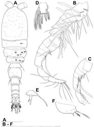
Male (based on allotype and one paratype from type locality). Total body length 448–473 µm (473 µm in allotype). Urosome with free genital somite. Habitus (Fig. 4A) more slender than in female, with prosome/urosome length ratio about 1.4 and greatest width in dorsal view at posterior end of cephalothorax. Body length/width ratio 3.7; cephalothorax about twice as wide as genital somite. Cephalothorax 1.3 times as long as wide (dorsal view); representing 35% of total body length. Ornamentation of cephalothorax (Figs 4A, 6A, B), free prosomites (Figs 4A, 6B), and first and last two urosomites (Fig. 5A, B, C) with same number and distribution of sensilla and pores as in female.
Genital somite (Fig. 5A, B, C) 1.5 times as wide as long in dorsal view, with serrated hyaline fringe dorsally, ornamented with one unpaired dorsal pore (no. 85), six pairs of dorsal and lateral sensilla (nos. 82, 86, 88-90), and three pairs of lateral pores (nos. 83, 84, 87); pores and sensilla nos. 82, 83, 87, 89 not present in female; no spermatophores visible inside. Third urosomite (Fig. 5A, B, C) homologous to posterior part of female genital double-somite, ornamented with dorsal unpaired pore (no. 91) and pores and sensilla nos. 92, 96, 97, but additionally with two pairs of lateral sensilla (nos. 93, 94) and one pair of lateral pores (no. 95). Fourth urosomite (Fig. 5A, B, C) similar to that of female, but ornamented with one additional pair of lateral sensilla (no. 101). Fifth urosomite (Fig. 5A, B, C) as in female. Anal somite (Fig. 5A, B, C) slightly shorter than in female, and with only one diagonal row of spinules on each side of anal sinus, but with other ornamentation and proportions of anal operculum as in female.
Caudal rami (Fig. 5A, B, C) slightly shorter than in female and with proportionally shorter innermost terminal seta, but with very similar ornamentation and armature to those of female; inner principal terminal seta with small constriction at start of pinnation.
Antennula (Figs 6F, 8B, C, D) strongly prehensile and digeniculate, 16-segmented (with ancestral 16th and 17th segments completely fused), ornamented with spinules only on first segment (as in female), with anvil-shaped cuticular ridges on anterior margin of 14th and 15th segments (distal geniculation). Armature formula as follows: 8+3ae.4.2.2+ae.2.2.2.2.2+ae.2.2.2.2 + ae.2.1+ae.11+ae. All aesthetascs linguiform and most relatively long and broad, apical one on 16th segment fused basally to one seta; most setae slender and smooth; short smooth setae on seventh (one) eighth (one), ninth (one), tenth (one), 12th (two), 13th (two), and 14th segments; short pinnate seta on 11th segment; six setae on 16th segment biarticulate distally or with breaking plane.
Antenna, labrum, mandibula, maxillula, maxilla, first swimming leg, second swimming leg, and third swimming leg as in female.
Fourth swimming leg (Fig. 4D) also with similar armature and ornamentation to that of female; third endopodal segment 1.8 times as long as wide; inner apical spine on third endopodal segment 1.3 times as long as outer apical spine and nearly as long as segment.
Fifth leg (Fig. 4E) similar to that of female, but with slightly shorter subapical exopodal spine.
Sixth leg (Fig. 4F) a large cuticular plate ornamented with single pore on anterior surface, armed with inner spine and two setae on outer distal corner; outermost seta unipinnate and 2.2 times as long as inner bipinnate seta, as well as 5.2 times as long as innermost spine.
Remarks
This species is probably most closely related to Diacyclops brevifurcus Ishida, 2006, which was described from Kyoto on the other side of Mount Hiei, in a parallel drainage basin to that of Lake Biwa (Ishida 2006[1]), and from Lake Biwa itself. The description of Diacyclops brevifurcus was quite lacking in detail, so we redecribed it after examining the holotype specimen dissected on one slide and four specimens from Lake Biwa (see below). Diacyclops brevifurcus shares with Diacyclops ishidai sp. n. the shape of the caudal rami, which are unusually short for the languidoides-group, and have an extremely large ventral protuberance on the distal margin with a pore on the tip. Both species also share exactly the same armature formula of all swimming legs as well as an identical pattern of cuticular pores and sensilla on all somites. Differences between the two include: relative length of the innermost terminal caudal seta, number of setae on the mandibular palp, relative length of the apical spine on the third leg endopod, number of rows of spinules on the intercoxal sclerite of the fourth leg, shape of the inner-distal margin of the fourth leg basis, and proportions of the distal segment of the fifth leg (all these arrowed in Figs 7, 8), as well as the armature of the antenna (with an exopodal seta present and only seven setae each on the second and third exopodal segments in Diacyclops brevifurcus). Ishida (2006)[1] illustrated two setae on the basis of the maxilliped in Diacyclops brevifurcus, but only one is present in the holotype (arrowed in Fig. 7F). Ishida (2002)[2] partly illustrated a specimen from Lake Biwa under the name Diacyclops sp. B, which he later (2006) attributed to Diacyclops brevifurcus (see the synonymy section below). We examined the four extant specimens from this series and agree that they are conspecific with Diacyclops brevifurcus (see below). Ishida erroneously illustrated the antenna of Diacyclops sp. B with only six setae on the second endopodal segment, and the antennula with only ten segments, but he later corrected himself in the description of Diacyclops brevifurcus.
Only four other species of the languidoides-group have similarly short caudal rami: Diacyclops ichnusae Pesce & Galassi, 1985 from Sardinia; Diacyclops ichnusoides Petkovski & Karanovic, 1997 from the ancient Lake Ohrid; Diacyclops improcerus (Mazepova, 1950) and Diacyclops versutus (Mazepova, 1962) from the ancient Lake Baikal (see Mazepova 1950[3], 1962; Pesce and Galassi 1985[4]; Petkovski and Karanovic 1997[5]). Unfortunately, all four were described based on a limited set of morphological characters, and many features cannot be compared with those of Diacyclops ishidai and Diacyclops brevifurcus. Five out of these six species come from or near ancient lakes, a distribution pattern commonly associated with Tertiary relics. Diacyclops ishidai has a shorter dorsal caudal seta and a more rounded seminal receptacle than Diacyclops ichnusae, longer caudal rami and third endopodal segment of the fourth leg than Diacyclops improcerus, a more slender habitus, longer female antennula, and shorter inner spine on the male sixth leg than Diacyclops versutus, and a shorter dorsal caudal seta, no exopodal seta on the antenna, and a longer third endopodal segment of the fourth leg than Diacyclops ichnusoides.
Original Description
- Karanovic, T; Grygier, M; Lee, W; 2013: Endemism of subterranean Diacyclops in Korea and Japan, with descriptions of seven new species of the languidoides-group and redescriptions of D. brevifurcus Ishida, 2006 and D. suoensis Ito, 1954 (Crustacea, Copepoda, Cyclopoida) ZooKeys, 267: 1-76. doi
Other References
- ↑ 1.0 1.1 Ishida T (2006) Diacyclops brevifurcus, a new cyclopoid copepod (Crustacea) from Mizoro-ga-ike pond, Kyoto city, and Lake Biwa, central Japan. Biogeography 8: 41-43.
- ↑ Ishida T (2002) Illustrated fauna of the freshwater cyclopoid copepods of Japan. Bulletin of the Biogeographical Society of Japan 57: 37-106.
- ↑ Mazepova G (1950) New species of cyclopoids in Lake Baikal. Doklady Akademii Nauk SSSR 75: 865-868. [in Russian]
- ↑ Pesce G, Galassi D (1985) Due nuovi Diacyclops del complesso “languidoides” (Copepoda: Cyclopidae) di acque sotterranee di Sardegna e considerazioni sul significato evolutivo dell’antenna nei copepod stigobionti. Bollettino del Museo Civico di Storia Naturale di Verona 12: 411-418.
- ↑ Petkovski T, Karanovic T (1997) Two new copepod species (Crustacea: Copepoda) from the Ohrid Lake. Annales de Limnologie 33: 245-253. doi: 10.1051/limn/1997023
Images
|
