The ripe tomatoes fruits present a good medium for the formation of many phytopathogenic and saprophytic microorganisms. One of such microorganisms, that is growing in the wide range of temperatures (4-35ºC), is the fungus Stemphylium botryosum Wallroth (Deuteromycotina).
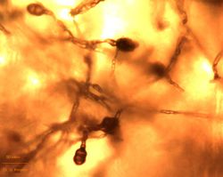
Infection is mainly through stomata and wounded surfaces of the plant. The fungus is confined to both leaves and fruits: the former show brownish spots, the latter – white-grayish mycelium that makes up a colony 1-2 cm diam. (fig. 1). It darkens quickly turning into dark gray, brownish and nearly black (fig. 2). Along with the surface mycelium from under the peel there develops a submersed one, which coming up on the surface form separate, not big, fascicular colonies consisting of the interlacement of mycelium hyphae and conidiophores with conidia (fig. 3, 4).
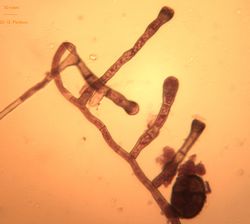
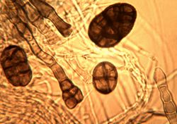
The mycelium hyphae are mainly colorless, but also can be yellowish-brown, 2-9 µm thick, with numerous septa (fig. 5, 6). Conidiophores, on which the conidia develop, are dark olive and dark brown, 10-80x3-7 µm, with apical bulging measuring 7-10 µm (fig. 7, 8). The septa of the conidiophores are placed 5-20 µm apart, but more often at a distance of about 10 µm. On the older conidiophores there often appear bulges (fig. 9, 10). The conidia are single, acanthaceous or verrucous, egg-shaped, roundish-square, occasionally of indefinite shape, with light ligatures near the septa and with a deeper ligature near the medium septa, yellowish or yellowish-olive in color, usually with 3-10 transverse and 1-10 longitudinal septa, 12-60x7-40 µm (fig. 11, 12).
Alongside with the tomato’s fruits the fungi can affect various plants, functions as a saprophyte or pathogen, infecting fruits, seed and leaves of Brassica oleracea, Brassica chinensis, Brassica juncea, Raphanus sativus, Allium cepa, Cucumis sativus, Daucus carota, Stachys sieboldii, Monarda didima, Hyssopus officinalis, Lophantus anisatus, Dracocephalum moldavica, Carum carvi, Coriandrum sativum, Foeniculum vulgare, Chrysanthemum сoronarium, Cichorium endivia, Physalis aequata, Lactuca sativa, Medicago sativa, Trifolium pretense, Phaseolus vulgaris, Pisum sativum and other agricultural cultures.
| 1.Tomato infected with the fungus Stemphylium botryosum (Image by G. Pestsov)
|
| 2.Stemphylium botryosum colony (Image by G. Pestsov)
|
| 3.Acanthaceous colonies of the fungus Stemphylium botryosum.JPG (Image by G. Pestsov)
|
| 4.Stemphylium botryosum mycelium, conidiophores and conidia.jpg (Image by G. Pestsov)
|
| 5.Formation of the conidia on the aerial mycelium of Stemphylium botryosum.jpg (Image by G. Pestsov)
|
| 6. Conidia formation on the aerial mycelium of Stemphylium botryosum.jpg (Image by G. Pestsov)
|
| 7.Young conidiphores formation.jpg (Image by G. Pestsov)
|
| 8.Conidiophores formation on the fungus mycelium.jpg(Image by G. Pestsov)
|
| 9.Conidiophore with a young conidium.JPG (Image by G. Pestsov)
|
| 10.Conidiophores and a conidium on the mycelium.JPG (Image by G. Pestsov)
|
| 11.Already formed acanthaceous conidia.JPG (Image by G. Pestsov)
|
| 12.The phases of conidiophores and conidia formation.JPG (Image by G.V. Pestsov)
|
|
| All images by Georgy Pestsov. |
(This page was moved here from http://phytopathology.net/Portal/Stemphylium_botryosum )


