Proarescon
| Notice: | This page is derived from the original publication listed below, whose author(s) should always be credited. Further contributors may edit and improve the content of this page and, consequently, need to be credited as well (see page history). Any assessment of factual correctness requires a careful review of the original article as well as of subsequent contributions.
If you are uncertain whether your planned contribution is correct or not, we suggest that you use the associated discussion page instead of editing the page directly. This page should be cited as follows (rationale):
Citation formats to copy and paste
BibTeX: @article{Huber2017JournalofHymenopteraResearch, RIS/ Endnote: TY - JOUR Wikipedia/ Citizendium: <ref name="Huber2017Journal of Hymenoptera Research">{{Citation See also the citation download page at the journal. |
Ordo: Hymenoptera
Familia: Mymaridae
Name
Proarescon Huber gen. n. – Wikispecies link – ZooBank link – Pensoft Profile
Type species
Borneomymar primitivum Huber, by present designation.
Diagnosis
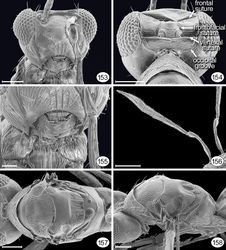
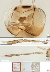
Female. Antenna with funicle 8-segmented (in Arescon 5-segmented) and clava 1-segmented, gradually narrowing apically to a point (Figs 156, 166). Both sexes. Fore wing with microtrichia more densely spaced except for oval area along posterior margin (in Arescon with microtrichia usualy more sparsely spaced, as shown in Triapitsyn [2016][1]).
Description
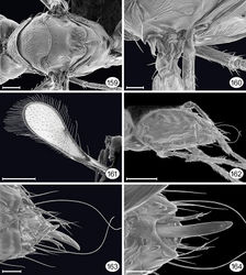
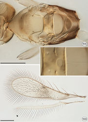
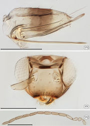
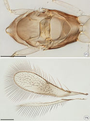
Female. Body 635–720 in length (critical point dried). Colour. Body generally light brown with some areas yellow to creamy white; darker brown are mouth margin, trabeculae, ocellar triangle, clava except apex, dorsellum, meso- and metapleuron, propodeum, and gt4–gt5 (Figs 165, 168, 170). Wings hyaline except for light brown behind venation (Fig. 169 and Huber 2002[2], fig. 5). Head. Head about 1.50–1.59× as wide as long, about 1.29–1.35× as wide as high, and 1.17–1.18× as high as long; in lateral view with anterior surface slightly convex, flat at level of toruli, then evenly curved to mouth margin; posterior surfaces convex and evenly curved from vertex to mouth margin. Face about 0.9× as wide as high; subantennal groove absent; preorbital groove ventral to level of torulus straight then more ventrally curving slightly medially to lateral margin of mouth opening (Figs 153, 171–male). Torulus in slight triangular depression about 1.7× as high as torulus width and separated by about 2.0× its width from transverse trabecula (Fig. 171–male). Vertex in lateral view horizontal, forming a right angle with face, posteriorly almost at right angle with occiput and separated from it by medially divided tranverse vertexal suture extending behind posterior ocelli almost from eye to eye; occiput separated from gena by a short, oblique posterior extension of supraorbital suture extending from lateral apex of vertexal suture and curving ventrally to dorsolateral corner of occipital foramen. Ocellar triangle small, slightly raised, with mid ocellus almost vertical and lateral ocelli oblique and facing posteriorly; ocelli with POL about 1.0× LOL and about 0.67× LOL; ocelli on stemmaticum (Fig. 154)—seen as white lines in cleared slide mounts (Fig. 165)—these are, respectively, a short, transverse groove in front of mid ocellus, continuing anterolaterally as the frontal suture to midpoint of supraorbital trabecula (apparently divided medially by an unscletotized area), a groove between the lateral margins of mid and lateral ocelli—the frontofacial suture, and a medially divided transverse groove behind the lateral ocelli, the vertexal suture, extending almost from eye to eye. Transverse trabecula darkly sclerotized medially and at each apex apparently not separated from supraorbital trabecula; preorbital trabecula extending ventrally about halfway between dorsal and ventral margins of torulus to where torulus nearest to eye; supraorbital trabecula in 2 almost equal sections, the anterior sections diverging posteriorly, the posterior sections parallel (Fig. 165). Eye large with numerous small facets, in lateral view about as high as wide and clearly separated dorsally from back of head (temple about 0.3× eye width). Ocular apodeme long and straight, needle-like. Malar sulcus present. Gena at level of ventral margin of eye slightly wider than malar space. Occiput separated from temple by occipital groove (Fig. 165) but otherwise not separated from gena/postgena. Mouthparts. Labrum with 1? seta; mandible with 4 uneven teeth (Fig. 153). Antenna. Scape 3.4–4.7× as long as wide, with radicle distinct from rest of scape and about 0.36–0.37× total scape length; pedicel about 2.0× as long as wide, almost as wide as and 0.26–0.27× as long as entire scape; funicle 8-segmented; clava 1-segmented, 0.98–1.10× as wide as apical funicle segment and 0.41–0.63× as long as entire funicle (Figs 156, 166, and Huber 2002[2], fig. 6). Mesosoma. About 1.7× as long as wide, 1.8× as long as high and 1.2× as wide as high. Pronotum entire (Fig. 157), in dorsal view clearly visible, medially about 0.5× as long as mesoscutum, with collar bell-shaped in lateral view pronotum sloping down towards junction with head and neck almost absent (not separable), and lateral panel somewhat rectangular and overlapping anterior margin of mesoscutum, with lateral surface merging smoothly into dorsal surface, with a shallow, oblique groove for femur. Spiracle (Fig. 157) flat with surface of pronotum, facing posterodorsally, and apparently slightly closer to anterior apex of notaulus than to posterolateral angle of pronotum. Propleura near anterior apex not quite abutting, then gap widening slightly more anteriorly. Prosternum rhomboidal and completely divided medially by faint longitudinal groove. Mesoscutum about 1.8× as long as scutellum, in dorsal view with shallow, thin, slightly diverging notauli a little wider and shallower posteriorly (Figs 157, 158, 168), in lateral view almost flat. Scutellum slightly wider than long, the anterior scutellum about 0.9× as long as frenum and separated from it by a shallow, medially straight frenal depression (Fig. 157); campaniform sensilla about as far from each other as to lateral margin of anterior scutellum; fenestra small, almost circular, and posterior to campaniform sensilla (Fig. 168, inset). Axilla distinctly advanced, the transscutal articulation laterally forming a distinct angle with median section (Fig. 168); axillula short, separated from anterior scutellum by concave axillular groove; mesophragma fairly narrowly convex posteriorly, extending to posterior apex of propodeum. Prepectus apparently narrowly triangular; mesopleuron somewhat rectangular, with shallow depression separating mesepisternum from mesepimeron. Metanotum with distinct triangular (Fig. 159) or lens-shaped (Fig. 168, in cleared slide mounts) dorsellum and lateral panel length toward hind wing articulation about one third length of dorsellum. Metapleuron triangular, the margin at junction with mesopleuron almost straight and posterior margin straight and vertical. Propodeum without carinae, with 1 propodeal seta (Fig. 159). Wings. Fore wing wide, with microtrichia on most of membrane beyond and partly behind venation to level of second macrochaeta except for a bare area medially along posterior margin (Fig. 169). Venation complete; submarginal vein with 1 proximal macrochaeta but no distal seta; parastigma 0.73× submarginal vein length; marginal vein present, its length about 1.42× parastigma length, with a second macrochaeta about midway between first distal macrochaeta and stigmal vein; stigmal vein distinct, about 0.28× length of marginal vein, with anterior margin of stigma parallel or converging with wing margin and with 4? apical campaniform sensilla in a line; postmarginal vein present, apparently about 1.1× as long and almost as thick as marginal vein; hypochaeta fairly close (about 0.3× length of parastigma) to proximal macrochaeta; proximal campaniform sensillum near posterior margin of parastigma just next to first distal macrochata. Hind wing normal (Fig. 169). Legs. Profemur slightly wider than meso-and metafemora; metafemur about 1.2× mesofemur width. Tarsi 5-segmented. Calcar (moveable protibial spur) with about 2 setae along outer margin, and with inner tine about 0.45× outer tine length. Middle and hind legs with tarsomere 1 as long as tarsomere 2. Metasoma. 1.95× as long as wide, 2.18× as long as high and 1.12× as wide as high; its length, excluding exserted part of ovipositor, about 1.37× that of mesosoma. Petiole ring-like, about 0.36× as long as wide. Gastral terga about equal in length except gt6 slightly longer (Figs 162, 170). Cercus flat, with 4 setae, the second-most dorsal one longest (Figs 162, 170). Hypopygium short, extending about one-third ovipositor length (Fig. 170). Ovipositor sheath exserted beyond gastral apex by about 0.2× total sheath length, with 1 subapical seta.
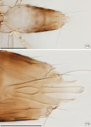
Male. Body length ≈ 585 (slide mounted paratype). Colour. Similar to female but with slightly more extensive brown on mesosoma (Fig. 173), and metasoma with brown apically instead of medially (Fig. 175). Head. As for female, mandible with 4 teeth (Fig. 171). Antenna. Scape (n=1) about 3.00× as long as wide, with radicle about 0.35× scape; pedicel 1.28× as long as wide; flagellum 11-segemented, with fl1 shorter and wider than other segments, fl2–fl11 subequal, each flagellomere with 4 mps (Fig. 172). Mesosoma. As for female (Figs 159, 160, 173). Wings. Fore wing (Fig. 174) with proximal campaniform sensillum near posterior margin of marginal vein about midway between first and second distal macrochata. Metasoma. Petiole length/width 0.38; gaster about 0.82× as long as mesosoma (Fig. 162). Genitalia with long parameres and apparently no digiti (Figs 163, 164, 176).
Etymology
The genus is masculine. The prefix, pro- is Latin for in front of, earlier or first, + Arescon, apparently the most closely related genus.
Key to species of Proarescon
Females.
Original Description
- Huber, J; 2017: Eustochomorpha Girault, Neotriadomerus gen. n., and Proarescon gen. n. (Hymenoptera, Mymaridae), early extant lineages in evolution of the family Journal of Hymenoptera Research, (57): 1-87. doi
Images
|
Other References
- ↑ Triapitsyn S (2016) Review of the Oriental species of the genus Arescon Walker, 1846 (Hymenoptera: Mymaridae). Euroasian Entomological Journal 15(Supplement 1): 137–151.
- ↑ 2.0 2.1 2.2 2.3 Huber J (2002) The basal lineages of Mymaridae (Hymenoptera) and description of a new genus, Borneomymar. In: Melika G Thuróczy C (Eds) Parasitic wasps. Evolution, systematics, biodiversity and biological control. Agroinform, Kiadó & Nyomba Kft., Budapest, 44–53.
