Phomopsis citri
Contents
Phomopsis citri H. Fawcett 1912
- non (Sacc.) Traverso & Spessa, hom. illeg. 1912
- = Phomopsis californica Fawcett, 1922.
- = Phomopsis caribaea Home, 1922.
- = Phomopsis cytosporella Penz. & Sacc., 1887.
Teleomorph:
- Diaporthe citri Wolf, Journal of Agricultural Research 33: 625, 1926 (as Diaporthe citri Fawcett).
Distribution
- Widespread in all Citrus-growing areas (CMI Map 126, ed. 2, 1966; Punithalingam & Holliday 1973 [1])
- Information on the host distribution in North-America is available at http://nt.ars-grin.gov/
Hosts
- On Citrus aurantifolia, C. aurantium, C. grandis, C. limon, C. parasisi, C. reticulata and C. sinensis.
Symptoms
Non-original description after Punithalingam & Holliday 1973 [1]:
Melanose of Citrus spp. and stem end rot of the fruit. Symptoms occur on the immature leaves, young branches, stalks and fruit. The very small spots enlarge, become water-soaked, sunken, dark with chlorotic halos and develop raised, corky, superficial, necrotic areas up to 1 mm diam.; this spotting is frequently very abundant and scar-like, necrotic aggregations are formed; on the fruit the spots are sometimes arranged in rings, lines or curves. Leaves are distorted and may fall prematurely. The small, dying branches bear the same raised spots in which both spore stages are found. Diaporthe citri is one of the citrus pathogens which penetrates the fruit at the stem end and causes a rot in storage.
Morphology
Non-original description after Punithalingam & Holliday 1973 [1]:
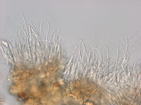
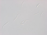
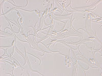
- Pycnidia on leaves, green and dead branches, bark and decaying fruit, scattered or clustered, immersed, later erumpent, black, conical to lenticular, up to 600 µm diam., with ostioles; wall many cells thick, composed of outer sclerotized cells and inner thin pseudoparenchyma lining the cavity. Conidiogenous cells enteroblastic, phialidic, hyaline, simple, cylindrical to obclavate, arising from the innermost layer of pseudoparenchymatic cells lining the pycnidial cavity. A-conidia (phialospores) hyaline, non-septate, fusiform to ellipsoidal, biguttulate, with one guttule at each end, 6-10 x 2-3 µm. B-conidia (phialospores) hyaline, non-septate, filiform, curved and often strongly hooked, 20-30 x 0.5-1 µm.
- Physiological specialization: None reported although differences in pathogenicity between isolates were found in Japan (50, 1238).
- Transmission: Water-borne through the conidia.
(End of data from CMI description)
Literature
- ↑ 1.0 1.1 1.2 Punithalingam, E. & Holliday, P. 1973, CMI Descriptions of Pathogenic Fungi and Bacteria 396.
- Wehmeyer, The Genus Diaporthe Nitschke and its segregates, 1933.
- Fawcett, Citrus diseases and their control, 1936.
- Knorr, Suit & Du Charme, Bulletin, Agricultural Experiment Stations, University of Florida 587, 1957.
- Atkinson, Diseases of tree fruits in New Zealand, 1971.
Strains studied
Strain BBA 72149
The strain was isolated from the wood of young Citrus branches after incubation in a moist chamber. It is listed here because the microscopical morphology of natural pycnidia on bark of Citrus is available. No DNA sequence exists for this exact strain, but a sequence of another Phomopsis strain sampled from the same tree (lab sequence number I3376). Furthermore, that sequence is identical with another strain isolated from Citrus from a neighboring tree, BBA 72156, for which both cultural measurements and DNA sequence are available (lab sequence number I3390; to be replaced with Genbank accession) - see below.
Observations of strain BBA 72149 from naturally infected material
Sequence identity with Ph. theicola!!!
Strain BBA 72156
The strain was isolated from the wood inside a twig. At a place where the bark and a small layer of wood was naturally ripped open, a elongate fruiting body An beschädigter Stelle eines Zweiges (1-15 cm,. dort abgespaltener Span) auf oder im Holz selbst (nicht Rinde) ein länglicher Fruchtkörper mit 3 Fruchthöhlen (Naturmaterial am 27.03. noch vorhanden)
Conidial measurements 51 day old (1% carrot juice agar, lab conditions):
- Alpha conidia: (6,3) Expression error: Unrecognized punctuation character ",".–Expression error: Unrecognized punctuation character ",". (8,5) × (2,1) Expression error: Unrecognized punctuation character ",".–Expression error: Unrecognized punctuation character ",". (2,8) µm [n = 18, mean / s. d. = 7,4 / 0,48 × 2,4 / 0,17]
- Beta conidia (measured along the curve or hook): (24,8) Expression error: Unrecognized punctuation character ",".–Expression error: Unrecognized punctuation character ",". (34,2) × (0,8) Expression error: Unrecognized punctuation character ",".–Expression error: Unrecognized punctuation character ",". (1,3) µm [n = 20, mean / s. d. = 30,2 / 2,5 × 1 / 0,13]
- Gamma conidia occasionally present.


