Ortheziola
| Notice: | This page is derived from the original publication listed below, whose author(s) should always be credited. Further contributors may edit and improve the content of this page and, consequently, need to be credited as well (see page history). Any assessment of factual correctness requires a careful review of the original article as well as of subsequent contributions.
If you are uncertain whether your planned contribution is correct or not, we suggest that you use the associated discussion page instead of editing the page directly. This page should be cited as follows (rationale):
Citation formats to copy and paste
BibTeX: @article{Kaydan2014ZooKeys406, RIS/ Endnote: TY - JOUR Wikipedia/ Citizendium: <ref name="Kaydan2014ZooKeys406">{{Citation See also the citation download page at the journal. |
Ordo: Hemiptera
Familia: Coccoidea
Name
Ortheziola Šulc, 1895 – Wikispecies link – Pensoft Profile
Type species
Ortheziola vejdovskyi Šulc, 1895, 1.
Diagnosis of genus
Adult female in life with a series of marginal, mediolateral and medial waxy protrusions, corresponding to wax plates on slide-mounted specimens. The distribution of these protrusions (and wax plates) differs between the species (Kozár 2004[1]).
Slide-mounted adult female with antenna 3-segmented; third antennal segment with slender apical seta, flagellate sensory seta and small subapical seta; second segment with 1 sensory pore. Eye stalk protruding, thumb-like, fused with sclerotized area at base of antenna, which is sometimes called the pseudobasal antennal segment. Legs well developed; leg setae robust, spine-like; trochanter and femur fused, tibia and tarsus fused; tibia with 1 sensory pore and at least 1 fleshy sensory seta; tarsus without digitule; claw digitules hair-like, claw without denticle. Labium 1-segmented, with many setae; with 3 long setae near apex of labium very close together, all situated in a single setal socket. Anal ring situated in a fold of derm on dorsal surface, ring bearing 6 setae. Sclerotized plate present on dorsum anterior to anal ring, wider than long. Modified pores, each with 2, 3 or 4 loculi, scattered over surface, appearing like microtubular ducts. Thumb-like pores forming cluster on each side of anal ring. Abdominal spiracles ventral on anterior segments, with at least one present on each side of segments I, II or III; when present, posterior abdominal spiracles located on dorsum near anal ring, surrounded by a cluster of multilocular pores (Kozár 2004[1]).
Distribution
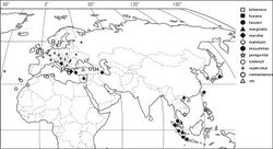
The 12 species of Ortheziola are distributed in the Palaearctic and North East part of the Oriental Regions (Figure 4). For detailed distribution data of the nine previously known species, see ScaleNet (Ben-Dov et al. 2013[2]). New locality records for several Ortheziola species were discovered during the study of the MHNG collection, which are listed below. The distribution patterns of the species may imply the existence of several other species in these regions, which would be worth further study.
Comments
The genus Ortheziola resembles the genera Ortheziolacoccus and Ortheziolamameti in having 3-segmented antennae, and the basal part of the antenna fused to the eye. However, Ortheziola differs from Ortheziolacoccus and Ortheziolamameti in having only a single spine band inside the ovisac band, and by its geographic distribution. The nine previously recorded species of Ortheziola occur in the Palaearctic and the North-East part of the Oriental Region.
Key to species of Ortheziola, based on adult females
Taxon Treatment
- Kaydan, M; Konczné Benedicty, Z; Szita, É; 2014: New species of the genus Ortheziola Šulc (Hemiptera, Coccoidea, Ortheziidae) ZooKeys, 406: 65-80. doi
Other References
- ↑ 1.0 1.1 Kozár F (2004) Ortheziidae of the World. Plant Protection Institute, Hungarian Academy of Sciences, Budapest, 525.
- ↑ Ben-Dov Y, Miller D, Gibson D (2013) Scalenet [accessed on 12 Dec 2013]. http://www.sel.barc.usda.gov/scalenet/scalenet.htm
Images
|
