Microporella appendiculata
| Notice: | This page is derived from the original publication listed below, whose author(s) should always be credited. Further contributors may edit and improve the content of this page and, consequently, need to be credited as well (see page history). Any assessment of factual correctness requires a careful review of the original article as well as of subsequent contributions.
If you are uncertain whether your planned contribution is correct or not, we suggest that you use the associated discussion page instead of editing the page directly. This page should be cited as follows (rationale):
Citation formats to copy and paste
BibTeX: @article{Martino2021ZooKeys1053, RIS/ Endnote: TY - JOUR Wikipedia/ Citizendium: <ref name="Martino2021ZooKeys1053">{{Citation See also the citation download page at the journal. |
Ordo: Cheilostomatida
Familia: Microporellidae
Genus: Microporella
Name
Microporella appendiculata (Heller, 1867) – Wikispecies link – Pensoft Profile
- Lepralia appendiculata Heller, 1867: 107, pl. 2, fig. 8.
- [[ | ]] ?Microporella coronata (Audouin & Savigny, 1826): Gautier 1962[1]: 173.
- Microporella coronata (Audouin & Savigny): Zabala 1986[2]: 513, fig. 180.
- Microporella marsupiata (Busk, 1860): Zabala 1986[2]: 514, fig. 181, pl. 15D.
- Microporella pseudomarsupiata Arístegui, 1984: 325, pl. 24, fig. 6; Zabala and Maluquer 1988[3]: 141, fig. 335, pl. 19C; Di Geronimo et al. 1993a[4]: table 1; Di Geronimo et al. 1997[5]: table 2; Chimenz and Faraglia 1995[6]: 40, table 1, pl. 2C; Rosso 1996a[7]: table 2.
- Microporella appendiculata (Heller): Hayward and Ryland 1999[8]: 294, figs 134A, B, 135 (cum syn.); Chimenz Gusso et al. 2014[9]: 187, fig. 100a–e.
Examined material
Italy • 2 living colonies; Ionian Sea, E Sicily, Ciclopi Island MPA; samples Ciclopi 2000 4E and 14G; 37°32'28"–37°34'30"N, 15°8'59"–15°11'1"E; 52 and 90 m; 16 Jul. 2000; A. Rosso leg.; dredging; DC and DL Biocoenoses; PMC Rosso-Collection I. H. B.84a. Italy • 27 living and 10 dead colonies/fragments; Ionian Sea, SE Sicily, Gulf of Noto; 36°41'45"–36°57'48"N, 15°8'35"–15°20'0"E; PS/81 cruise; samples CR1, 9B and 10C; 45, 44 and 60 m; Jul. 1981; I. Di Geronimo leg.; dredging; DC Biocoenoses; and 3 living colonies; Noto 1996 cruise; samples 6C and 9E; 45–50 m; 1996; E. Mollica leg.; dredging; VTC and DC Biocoenoses; PMC Rosso-collection I. H. B84c. Italy • 5 living colonies; Iberian-Provençal Basin, NW Sardinia, Capo Caccia-Punta Giglio MPA; samples Bisbe 1, Bisbe 2 and Falco 1; 40°35'40"N, 8°11'39"E; 7–8 m; Jun. 2009; V. Di Martino leg.; submarine cave; scuba diving; PMC Rosso-Collection I. H. B.84b. France • 11 dead colonies; Iberian-Provençal Basin, Corsica, off Calvì; sample CL 74; 42°47'31"N, 9°8'10"E; 150–110 m; G. Fredj leg.; dredging; DL Biocoenosis; PMC Rosso-collection Fr. H. B84d. Greece • 1 dead colony; NE Aegean Sea, Lesvos Island, Agios Vasilios cave; sample AV1; 38°58'9"N, 26°32'28"E; ca. 30 m, V. Gerovasileiou leg.; submarine cave; scuba diving; PMC Rosso-collection Gr. H. B84e.
Description
Colony encrusting multiserial, unilaminar, forming subcircular patches; interzooidal communications typically via two proximolateral, two distolateral and three distal pore-chamber windows, 48–122 (71±25, N = 10) × 16–26 μm (20±3, N = 10) along lateral walls.
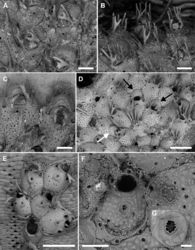
Autozooids polygonal, 529–742 (644±66, N = 14) × 347–582 (458±66, N = 14) μm (mean L/W = 1.41), distinct, the boundaries marked by narrow grooves between the slightly raised vertical walls (Fig. 2D, E). Frontal shield flat to slightly convex, coarsely, densely and evenly granular; 5–8 marginal areolae only occasionally distinguishable from pseudopores; pseudopores circular to elliptical (6–16 μm in diameter), numbering 30–42 (fewer in periancestrular zooids), placed in the proximal half of the zooid (Fig. 2E); area between orifice and ascopore imperforate.
Primary orifice transversely D-shaped, 100–110 (105±7, N = 2) × 129–141 μm (135±8, N = 2) (mean OL/OW = 0.78; mean ZL/OL = 6.14); hinge-line straight or concave, smooth, without condyles and denticles. Five, occasionally six, articulated oral spines, 170–310 μm long (diameter of the base 25–42 μm), the proximalmost pair bi- to trifurcated, the tips sometimes curved towards the centre of the orifice (Fig. 2A, B); joints brown.
Ascopore field an area of smooth, gymnocystal calcification, placed 50–80 μm below the orifice, transversely elliptical, 48–122 × 61–110 μm, narrow distally, more extensive proximally and developing a prominent, pointed mucro not concealing the ascopore; ascopore subcircular, 22–43 μm in diameter, with a dozen of radial spines (Fig. 2F, G).
Avicularia paired, 72–170 (103±29, N = 37) × 46–103 (67±13, N = 37) μm (mean AvL/AvW = 1.53), located distolaterally, the complete crossbar at the same level of the orifice hinge-line (Fig. 2D, E); rostrum short, arched or truncated distally and open-ended, distally directed. Mandible brown, 387–659 μm long, setiform, typically curved, lying on the distal zooid (Fig. 2A).
Ovicell subglobular and semi-immersed, 143–235 (195±29, N = 15) × 262–378 (329±38, N = 15) μm (mean OvL/OvW = 0.59), non-personate, not obscuring the proximal part of the orifice, closed by the operculum during brooding (Fig. 2A, C, D), in the same colony either kenozooidal (Fig. 2C, D black arrows) or produced by the distal autozooid (Fig. 2D white arrow); surface finely granular, imperforate; a proximal rim of gymnocystal calcification occupying about one-third of the ooecium length, forming a raised visor-like rim; proximalmost pair of spines (branched) visible in ovicellate zooids.
Ancestrula tatiform (Fig. 2E), oval (424 × 324 μm), gymnocyst more extensive proximally than laterally, about 150 μm wide; opesia pyriform (270 × 180 μm), surrounded by a smooth, flared cryptocyst with the undulate marginal rim indented by ten gymnocystal spines (six distal, two median, three proximal). Ancestrula budding two distolateral autozooids, often regenerated as a small autozooid lacking avicularia (Fig. 2F).
Remarks
Originally described from the Adriatic by Heller (1867)[10], Microporella appendiculata has often been recorded as Microporella marsupiata Busk, 1860 or as M. pseudomarsupiata Arístegui, 1984 (see Hayward and Ryland 1999[8] and references therein; Rosso et al. 2010[11]; Chimenz Gusso et al. 2014[9]). With the exception of Hayward and Ryland (1999)[8] and recent online material such as the catalogue of the Museu de Ciènces Natural de Barcelona (https://zoologiaenlinia.museuciencies.cat/detall/zoologia_general/N1064271/) and the website of the program DORIS (https://doris.ffessm.fr/Especes/Microporella-pseudomarsupiata-Microporelle-ciliee-noire-1695), a comprehensive description and illustration of this species is absent in the literature.
Microporella flabelligera Levinsen, 1909 described from the vicinity of Siracusa, at depths (28–46 m) comparable to those of the PS/81 sites in the Gulf of Noto, is likely to be conspecific with M. appendiculata based on the original drawings (Levinsen 1909[12]: 331, pl. 24 cited as 23 in the text, fig. 6A–C). However, a formal synonymy requires the examination of the type specimen.
Microporella appendiculata differs from other Mediterranean congeners in having paired avicularia, a character shared only with M. coronata (Audouin & Savigny, 1826). However, in M. coronata the avicularia are located proximally to the ascopore, the ovicell is personate, the oral spines are greater in number (6–8) and unbranched.
Here, we document the regeneration of the ancestrula as an autozooid for the first time (Fig. 2F). Trifurcated proximal spines have already been figured on some zooids, though not described as such by Zabala (1986[2]: fig. 181B, as M. marsupiata) and by Chimenz and Faraglia (1995[6]: pl. 2C, as M. pseudomarsupiata). Gautier (1962)[1] also recorded autozooids with up to seven oral spines.
Size differences were observed between specimens from Sicily and those from Sardinia, with Sardinian colonies showing longer autozooids (Sicily: mean 604±56 × 458±71 μm, N = 8, L/W 1.32; Sardinia: 698±30 × 458±64 μm, N = 6, L/W 1.53), slender avicularia (Sicily: mean 135±7 × 10⁵±8 μm, N = 25, L/W 1.38; Sardinia: 142±14 × 80±14 μm, N = 12, L/W 1.78), and larger ovicells (Sicily: mean 173±20 × 280±14 μm, N = 5, L/W 0.78; Sardinia: 206±27 × 354±13 μm, N = 10, L/W 0.58), as well as trifurcated proximalmost spines.
Distribution and ecology
In the Mediterranean, M. appendiculata has been reported from several localities and usually in shelf habitats, associated with coarse detritic bottoms, often encrusting shells and calcareous algae (Gautier 1962[1]; Rosso 1996a[7], b[13]; Madurell et al. 2013[14]; Chimenz Gusso et al. 2014[9]), as well as from shadowed microhabitats associated with Cystoseira s. l. communities (e.g., Campisi 1973[15]; Rosso et al. 2019a[16]). It also occurs in submarine caves of NW Sardinia (Fraschetti et al. 2010[17]) and near Monaco (J.-G. Harmelin, pers. comm., May 2021) in the Iberian-Provençal basin, at the transition between coralligenous and semi-dark cave habitats. Microporella appendiculata has been also collected in dark sectors of Agios Vasilios cave in Lesvos, Greece (Rosso et al. 2019b[18]) and (as M. umbracula) in the Mitigliano cave in the Sorrento peninsula, Italy (Balduzzi et al. 1989[19]; Balduzzi and Rosso 2003[20]).
Taxon Treatment
- Martino, E; Rosso, A; 2021: Seek and ye shall find: new species and new records of Microporella (Bryozoa, Cheilostomatida) in the Mediterranean ZooKeys, 1053: 1-42. doi
Images
|
Other References
- ↑ 1.0 1.1 1.2 Gautier Y (1962) Recherches ecologiques sur les Bryozoaires Chilostomes en Méditerranée occidentale.Recueillis des Travaux de la Station Marine d’Endoume38: 1–435.
- ↑ 2.0 2.1 2.2 Zabala M (1986) Fauna dels bryozous dels Països Catalans. Barcelona.Institut D’estudis Catalans Secció de Ciències84: 1–836.
- ↑ Zabala M, Maluquer P (1988) Illustrated keys for the classification of Mediterranean Bryozoa.Treballs del Museu de Zoologia, Barcelona4: 1–294.
- ↑ Di Geronimo I, La Perna R, Rosso A, Sanfilippo R (1993a) Popolamento e tanatocenosi bentonica della Grotta dell’Accademia (Ustica, Mar Tirreno meridionale), Naturalista Siciliano (Serie 4) 17(1–2): 45–63.
- ↑ Di Geronimo I, Allegri L, Improta S, La Perna R, Rosso A, Sanfilippo R (1997) Spatial and temporal aspects of Recent benthic thanatocoenoses in a Mediterranean Infralittoral cave.Rivista Italiana di Paleontologia e Stratigrafia103(1): 15–28.
- ↑ 6.0 6.1 Chimenz C, Faraglia E (1995) Some faunistic and ecological observations on the Bryozoa Gymnolaelata assemblages from the coast of Puglia (Italy). Atti della Società Toscana di Scienze Naturali, Memorie, ser.B102: 37–47.
- ↑ 7.0 7.1 Rosso A (1996a) Popolamenti e tanatocenosi a Briozoi di fondi mobili circalitorali del Golfo di Noto (Sicilia SE). Naturalista Siciliano (Serie 4) 20(3–4): 189–225.
- ↑ 8.0 8.1 8.2 Hayward P, Ryland J (1999) Cheilostomatous Bryozoa. Part 2. Hippothoidea – Celleporoidea. Barnes RSK, Crothers JH (Eds) Synopses of the British Fauna (New Series) Field Studies Council, Shrewsbury 14: 1–416.
- ↑ 9.0 9.1 9.2 Chimenz Gusso C, Nicoletti L, Bondanese C (2014) Briozoi. Biologia Marina Mediterranea 20 (Suppl. 1): 1–336.
- ↑ Heller C (1867) Die Bryozoen des adriatischen Meeres.Verhandlungen der zoologisch-botanischen Gesellschaft in Wien17: 77–136.
- ↑ Rosso A, Chimenz Gusso C, Balduzzi A (2010) Bryozoa. In: Relini G (Ed.) Checklist della flora e della fauna dei mari italiani (parte II).Biologia Marina Mediterranea 17(Suppl.), 589–615.
- ↑ Levinsen G (1909) Morphological and Systematic Studies on the Cheilostomatous Bryozoa.Nationale Forfatterers Forlag, Copenhagen, 431 pp. https://www.biodiversitylibrary.org/page/4713708
- ↑ Rosso A (1996b) Valutazione della biodiversità in Mediterraneo: l’esempio dei popolamenti a briozoi della Biocenosi del Detritico Costiero.Biologia Marina Mediterranea3(1): 58–65.
- ↑ Madurell T, Zabala M, Dominguez-Carrió C, Gili J (2013) Bryozoan faunal composition and community structure from the continental shelf off Cap de Creus (Northwestern Mediterranean).Journal of Sea Research83: 123–126. https://doi.org/10.1016/j.seares.2013.04.013
- ↑ Campisi M (1973) Briozoi dell’Isola Lachea (Golfo di Catania) – Nota preliminare. Bollettino Accademia Gioenia di Scienze Naturali, Catania (Serie 4) 11: 135–156.
- ↑ Rosso A, Sanfilippo R, Sciuto F, Serio D, Catra M, Alongi G, Viola A, Leonardi R (2019a) Preliminary information on bryozoans associated with selected Cystoseira communities from Sicily (Mediterranean). In: Schmidt R Reid C Gordon D Walker‐Smith G Martin S Percival I (Eds) Bryozoan Studies 2016. Proceedings of the 17th International Bryozoology Association Conference, Melbourne, Australia.Memoirs of the Australasian Association of Palaeontologists52: 115–129.
- ↑ Fraschetti S, Boero F, Guarnieri G, Terlizzi A, Guidetti P, Bussotti S, Piraino S, De Vito D, Cormaci M, Furnari G, Catra M, Alongi G, Rosso A, Di Martino E, Ceccherelli G, Manconi R, Ledda F, Cattaneo-Vietti R, Cerrano C, Pantaleo U, Scinto A, Bavestrello G, Di Camillo C, Betti F, Chemello R, Milazzo M, Graziano M, Di Franco A, Marchini A, Russo G, Di Stefano F, Cimmino P (2010) Studio degli ambienti di grotte marine sommerse (Cod.8330) nelle aree marine protette di Capo Caccia, Plemmirio e Isole Pelagie. Relazione finale. CoNISMa-Ministero dell’Ambiente e della Tutela del Territorio e del Mare, 337 pp.
- ↑ Rosso A, Gerovasileiou V, Sanfilippo R, Guido A (2019b) Bryozoans assemblages from two submarine caves in the Aegean Sea (Eastern Mediterranean).Marine Biodiversity49(2): 707–726. https://doi.org/10.1007/s12526-018-0846-0
- ↑ Balduzzi A, Bianchi C, Boero F, Cattaneo Vietti R, Pansini M, Sarà M (1989) The suspension-feeder communities of a Mediterranean sea cave. In: Ros J (Ed.) Topics in Marine Biology.Scientia Marina53: 387–395.
- ↑ Balduzzi A, Rosso A (2003) Briozoi. In: Cicogna F, Bianchi NC, Ferrari G, Forti P (Eds) Grotte marine: cinquant’anni di ricerca in Italia, CLEM-ONLUS, Ministero dell’Ambiente e della Tutela del Territorio: 195–202.
