Macandrewella cochinensis
| Notice: | This page is derived from the original publication listed below, whose author(s) should always be credited. Further contributors may edit and improve the content of this page and, consequently, need to be credited as well (see page history). Any assessment of factual correctness requires a careful review of the original article as well as of subsequent contributions.
If you are uncertain whether your planned contribution is correct or not, we suggest that you use the associated discussion page instead of editing the page directly. This page should be cited as follows (rationale):
Citation formats to copy and paste
BibTeX: @article{El-Sherbiny2013ZooKeys344, RIS/ Endnote: TY - JOUR Wikipedia/ Citizendium: <ref name="El-Sherbiny2013ZooKeys344">{{Citation See also the citation download page at the journal. |
Ordo: Calanoida
Familia: Scolecitrichidae
Genus: Macandrewella
Name
Macandrewella cochinensis Gopalakrishnan, 1973 – Wikispecies link – Pensoft Profile
Material examined
Nine adult females and eight adult males collected from Sharm El-Maya Bay located in the entrance of Sharm El-Sheikh City, the northern Red Sea on 5 December 2011.
Body length. Female: 2.88–3.15 mm (mean±SD=2.99±0.09 mm, n=6). Male: 2.83–3.21 mm (2.98±0.13 mm, n=6).
Female
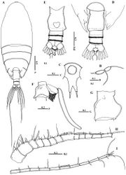
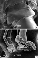
Body (Fig. 2A) robust; cephalosome completely fused to first pediger, protruding anteroventerally into bifurcated rostrum; rostrum (Figs 2B–C) with pair of slender filaments; single median cuticular lens present at base of rostrum (Figs 2B–C, 3A). Pedigers 4 and 5 partially fused, with incomplete suture visible dorsally and ventrolaterally; posterior margin asymmetrical, left one longer; each with 1 pairs of processes on each side, postero-dorsolateral projecting on each side lamellar with serrated margin, asymmetrical ventrolateral processes curved ventromedially at tip, slightly exceeding the posterior end of genital double somite on left side and slightly exceeding half length of genital double-somite on right (Figs 2D, 3B, D). Urosome (Figs 2E, D) short, approximately one-fifth as long as prosome; of 4 free somites. Genital double-somite asymmetrical with unequal anterodorsal protrusion on each side and posterodorsal swelling on left side (Figs 2F, G, 3C, D); genital area usually with sausage-like spermatophore (Fig. 2F); genital operculum wider than long, located distoventrally (Fig. 2E). Fourth urosomite (anal somite) very short, telescoped into proceeding somite. Caudal rami symmetrical with 5 caudal setae, left middle seta (V) 1.5 times as long as right one. Antennules (Figs 2H, I) symmetrical, 23-segmented, extending nearly to posterior border of second somite. Segmentation pattern and setal armature elements as follows: I-3, II-IV, 6+ae (II-2, III-2+ae, IV-2), V-2+ae, VI-2, VII-1+ae, VIII-2, IX-2+ae, X-XII-4+ae, XIII-1, XIV--2+ae, XV-1, XVI-2+ae, XVII-1, XVIII-1, XIX-1, XX-2, XXI-1+ae, XXII-1, XXIII-1, XXIV-1+1, XXV-1+1, XXVI-1+1, XXVI-XXVIII-5+ae.
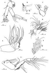
Antenna (Fig. 4A) coxa with 1 plumose seta medially and lateral array of curved setules; basis with 2 mediodistal setae of unequal length. Exopod 7-segmented with setal formula of 0, 0-0-1, 1, 1, 1, 1, 1+3 setae; endopod 2-segmented, first segment with 2 subterminal setae and patch of fine setules medially, distal segment bearing 8 setae on middle lobe, terminal lobe with 7 setae and patch of fine setules. Mandible gnathobase (Fig. 4B) heavily sclerotized with cutting edge bearing 8 teeth (5 of them flattened with broad edge) and spinulose seta. Palp (Fig. 4C) basis longer than wide, bearing 2 spinulose setae; exopod consisting of 5 segments with setal formula of 1, 1, 1, 1, 2; endopod 2-segmented, with 2 setae on first segment and 9 setae and row of fine spinules on second segment.
Maxillule (Fig. 4D) with praecoxal arthrite bearing 13 setae, 9 setae along terminal border, 4 setae on posterior surface and 1 seta on anterior surface (Fig. 4D). Coxal endite bearing 2 setae; coxal epipodite with 9 setae; basis completely fused with endopod; first and second basal endites with 3 and 5 setae respectively; baseoendopod with 7 setae terminally; exopod lobate, bearing 8 setae.
Maxilla (Figs 4E, F) praecoxal endite 1 with 4 setae, second praecoxal to second coxal endites each bearing 3 setae; basis with 2 setae and 2 worm-like sensory setae and patch of fine spinules. Endopod (Figs 3E, 4F) indistinctly three-segmented, bearing 3 brush-like, 2 brush-like and 3 worm-like sensory setae, respectively.
Maxilliped (Fig. 4G) praecoxal endites of syncoxa with 2 worm-like and 1 hirsute setae proximally, and 1 brush-like setae at nearly mid-length; coxal endite with 3 setae located at distal end. Basis nearly as long as syncoxa with submarginal row of minute spinules and 3 setae along medial margin. Endopod 6-segmented; first endopodal segment very short and almost incorporated into basis bearing 2 setae; second to sixth endopodal segment with setal formula of 4, 4, 3, 3+1, 4.
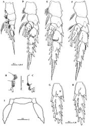
Legs 1 to 4 biramous, with 3-segmented exopods; endopod 1-segmented in leg 1, 2-segmented in leg 2, 3-segmented in legs 3 and 4. Spines and setal formula are shown in Table 1. Leg 1 (Figs 5A–C) smallest, first exopodal segment with expanded medial margin bordered by naked lateral spinules (Fig. 5B), middle segment bearing lateral spine and medial seta, distal exopod segment with serrate spine and spiniform terminal seta; endopod bearing middle lateral knob with patch of fine setules terminally (Fig. 5C). Leg 2 (Fig. 5D) coxa and basis with pointed prominence on lateral margin; second exopodal segment with crescent-like row of spinules on posterior surface; third segment with middle patch of spinules posteriorly; first endopodal segment without any spinules; second endopodal segment bearing 6 acute spinules. Leg 3 (Fig. 5E) coxa with pointed prominence on lamellar lateral margin; basis with pointed process on medial distal corner; second exopodal segment with crescent-like row of spinules along distal margin, third segment with minute spinules distributed in curved row; second and third endopodal segments bearing 4 and 6 spinules, respectively. Leg 4 (Fig. 5F): second and third exopodal segments each bearing longitudinal row of stout spinules distributed as shown in Fig. 5F. Shape, number and distribution of spinules along second and third exopodal segment varies among individuals (Figs 5G, H).
| Exopod | Endopod | |||||||
|---|---|---|---|---|---|---|---|---|
| Coxa | Basis | 1 | 2 | 3 | 1 | 2 | 3 | |
| Leg 1 | 0-0 | 0-1 | I-0; | I-1; | I,1,3 | 0,2,3 | ||
| Leg 2 | 0-1 | 0-0 | I-1; | I-1; | III,I,4 | 0-1; | 1,2,2 | |
| Leg 3 | 0-1 | 0-0 | I-1; | I-1; | III,I,4 | 0-1; | 0-1; | 1,2,2 |
| Leg 4 | 0-1 | 0-0 | I-1; | I-1; | III,I,4 | 0-1; | 0-1; | 1,2,2 |
Male
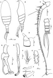
Body (Figs 6A, B) more slender than female; rostrum bifurcated with pair of filaments; cuticular median lens present at base of rostrum. Cephalosome completely fused with first pediger, fourth and fifth pedigers fused with suture visible laterally; border of fifth pediger symmetrical, ending with paired stout ventrally-curved processes. Urosome (Fig. 6C) 5-segmented; genital somite asymmetrical, with anterior dorsal knobs on right side (Figs 6C, D, 7A); second to fourth urosomites with thin spinules along posterior margin; second urosomite slightly asymmetrical in dorsal view, anal somite very small; caudal rami symmetrical, each ramus bearing 4 plumose setae. Antennule (Fig. 5E) consisting of 18 and 19 articulated segments on right and left side, respectively. Setal formula of left antennule as follows: I-1+ae, II-IV-6+4ae (II-2+ae, III-2+2ae, IV-2+ae), V-2+2ae, VI-2+ae, VII-2 (1 missed)+2ae, VIII-2+ae, IX-2+2ae, X-XV-7+6ae, XVI-XVII-2+3ae, XVIII-1+ae, XIX-1+ae, XX-1+ae, XXI-1+ae, XXII-unarmed, XXIII-1, XXIV-1(missed)+ae, XXV-1+1, XXVI-1+1, XXVII-XXVIII-5+ae. Right antennules of 18 free segments with fusion of segments XXII and XXIII; setal formula of I-1+ae, II-IV-6+4ae, V-2+2ae, VI-2+ae, VII-1+ae, VIII-2+ae, IX-2+2ae, X-XV-5+6ae, XVI-XVII-2+3ae, XVIII-1+ae, XIX-1+ae, XX-1+ae, XXI-1+ae, XXII-XXIII-1, XXIV-1+1+ae, XXV-1+1+ae, XXVI-1+1, XXVII-XXVIII-5+ae.
Mouth parts and legs 1-4 similar to those of female except fifth and sixth endopodal segment of maxilliped with longer setae (Fig. 5F) and third exopodal segment of leg 2 with different number and distribution of posterior surface setules (Fig. 5G).
Leg 5(Figs 6H–K) elongated in general structure resembling that of the other species of the genus. Left leg (Fig. 6H) with coxa approximately as long as basis; basis with longitudinal keel–like structure along proximal half; exopod 2–segmented, second segment with lamellar plate covered with dense tuft of cilia and 2 elements terminally (Figs 6I, 7B); endopod one-segmented, shorter than exopod, bearing 2 medial triangular processes, one seta at tip and medially serrated margin (Fig. 6J). Right leg chelate (Fig. 6K); coxa with triangular expansion proximally; basis expanded laterally; first exopodal segment bearing 3 medial processes, one located proximally, middle irregular and distal somewhat triangular; second exopodal segment short, bearing internally directed process truncate curved at tip; third segment as long as previous segment, curved inward distally (Fig. 7B); endopod one-segmented, curved outward and recurved at tip, bearing round process distally and triangular process midway.
Taxon Treatment
- El-Sherbiny, M; Al-Aidaroos, A; 2013: First record and redescription of Macandrewella cochinensis Gopalakrishnan, 1973 (Copepoda, Scolecitrichidae) from the Red Sea, with notes on swarm formation ZooKeys, 344: 1-15. doi
Images
|
