Liminympha makarkini
| Notice: | This page is derived from the original publication listed below, whose author(s) should always be credited. Further contributors may edit and improve the content of this page and, consequently, need to be credited as well (see page history). Any assessment of factual correctness requires a careful review of the original article as well as of subsequent contributions.
If you are uncertain whether your planned contribution is correct or not, we suggest that you use the associated discussion page instead of editing the page directly. This page should be cited as follows (rationale):
Citation formats to copy and paste
BibTeX: @article{Makarkin2013ZooKeys325, RIS/ Endnote: TY - JOUR Wikipedia/ Citizendium: <ref name="Makarkin2013ZooKeys325">{{Citation See also the citation download page at the journal. |
Ordo: Neuroptera
Familia: Nymphidae
Genus: Liminympha
Name
Liminympha makarkini Ren & Engel, 2007 – Wikispecies link – Pensoft Profile
- Liminympha makarkini Ren & Engel, 2007:212, figs 1–3; Engel and Grimaldi 2008[1]:9; Yang et al. 2010[2]:177.
Redescription
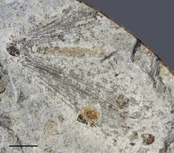
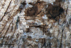
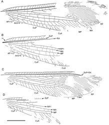
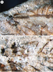
Body (metathorax, abdomen) poorly, fragmentarily preserved. First abdominal tergite rather long; distally with distinct transverse suture, probably heavily sclerotized; medially with mediolongitudinal short suture; portion of 1st tergite distal to transverse suture (‘Transversalnaht’ of Achtelig 1975[3]) very narrow (Fig. 4). Second tergite nearly as wide as long. Other tergites indistinct. Forewing elongate, narrowed in proximal portion, most dilated at distal 3/4 length; about 30 mm long, 8 mm wide. Costal space narrow, basally narrowed, distally dilated. All preserved subcostal veinlets simple, strongly oblique distally; veinlets of ScP+RA dichotomously branched. Humeral veinlet rather strongly recurrent, branching not detected (Fig. 6B). Subcostal space very narrow; two crossveins in distal part detected, others possible. RA spaces slightly narrower than costal space, narrowed towards apex; crossveins irregularly spaced for entire preserved portion. RP originates rather near wing base, with about 16 branches; RP1 originates far from origin of RP; at least four proximal-most branches widely spaced, more distal branches closely spaced. In right forewing, RP2 terminated at RP1; RP3, RP4 fused for short distance (probably aberrations). Crossveins between branches of RP very scarce, restricted to area between RP1 to RP5. M appears fused with R basally; forked at nearly equal distance from origin of RP, origin of RP1. MA long, slightly arched, distally few-branched. MP long, its anterior trace (stem of MP) nearly straight before terminal branching; with five long distal branches, quite strongly inclined. Crossveins between R/RP and MA, MA and MP irregularly spaced, arranged differently in right, left wings. Cu dividing into CuA, CuP rather near to wing base. CuA long, smoothly curved anteriorly, pectinate with four long branches, each dichotomously branched. Between branches of CuA at last three crossveins forming gradate series continued CuP (‘pseudo-CuP’). CuP long, pectinately branched, with seven-eight rather short branches, most simple. AA3+4 rather short; stem simple, with one or two simple branches. AP1+2 incompletely preserved, probably simple. AP3+4 not preserved. One dark rather big spot in distal portion of radial space might be present (apical half of other wing not preserved). Hind wing similar in shape to forewing, slightly narrower; about 28 mm long, 7 mm wide. Costal space narrow, dilated distally. Subcostal veinlets simple, becoming more oblique, closely spaced, curved towards apex. Humeral veinlet bent to base, nearly straight, not branched. Subcostal space narrow; three crossveins detected, others possible. ScP, RA fused far from wing apex; preserved veinlets of ScP+RA long, dichotomously forked; crossveins between them not detected. RA space basally as wide as costal space, narrowed towards apex; crossveins rather regularly spaced for entire preserved portion (right wing). RP originated near wing base, with about 15 branches; RP1 originated far from origin of RP (but closer to wing base than in forewing); five proximal-most branches widely spaced, other (distal) branches closely spaced. Crossveins between branches of RP very scarce, restricted to area between RP1 to RP4. Origin of M and its fork not preserved. MA long, nearly straight, distally dichotomously branched. MP long, its anterior trace (stem of MP) slightly incurved, with four long distal branches, quite strongly inclined. Origin of Cu and its dividing into CuA, CuP not present. CuA long, pectinate, with 15 branches, which proximally simple, distally once or twice forked. CuP short, deeply forked. Distal crossvein between CuA, CuP rather short connecting CuA, anterior branch of CuP fork. Anal veins not preserved. Crossveins between R/RP and MA, MA and MP, MP and CuA poorly preserved, irregularly spaced, arranged differently in right and left wings; crossveins between branches of MP, CuA absent.
Material
Holotype CNU-NEU-NN1999024 (part, counterpart), deposited in CNUB; an incomplete specimen.
Type locality and horizon
Daohugou Village, Shantou Township, Ningcheng County, Inner Mongolia, China. Jiulongshan Formation, Middle Jurassic.
Remarks
This redescription is only based on the counterpart of the holotype.
Taxon Treatment
- Makarkin, V; Yang, Q; Shi, C; Ren, D; 2013: The presence of the recurrent veinlet in the Middle Jurassic Nymphidae (Neuroptera): a unique character condition in Myrmeleontoidea ZooKeys, 325: 1-20. doi
Other References
- ↑ Engel M, Grimaldi D (2008) Diverse Neuropterida in Cretaceous amber, with particular reference to the paleofauna of Myanmar (Insecta). Nova Supplementa Entomologica 20: 1-86.
- ↑ Yang Q, Ren D, Shih C, Wang Y, Shi C, Peng Y, Zhao Y (2010) Neuroptera – Grace with Lace. In: Ren D Shih C Gao T Yao Y Zhao Y. Silent Stories – Insect Fossil Treasures from Dinosaur Era of the Northeastern China. Sceince Press, Beijing, 165–179.
- ↑ Achtelig M (1975) Die Abdomenbasis der Neuropteroidea (Insecta, Holometabola). Eine vergleichend anatomische Untersuchung des Skeletts und der Muskulatur. Zoomorphologie 82: 201-242. doi: 10.1007/BF00993588
Images
|


