Kijanebalola devestiva
| Notice: | This page is derived from the original publication listed below, whose author(s) should always be credited. Further contributors may edit and improve the content of this page and, consequently, need to be credited as well (see page history). Any assessment of factual correctness requires a careful review of the original article as well as of subsequent contributions.
If you are uncertain whether your planned contribution is correct or not, we suggest that you use the associated discussion page instead of editing the page directly. This page should be cited as follows (rationale):
Citation formats to copy and paste
BibTeX: @article{Todaro2013ZooKeys315, RIS/ Endnote: TY - JOUR Wikipedia/ Citizendium: <ref name="Todaro2013ZooKeys315">{{Citation See also the citation download page at the journal. |
Ordo: Chaetonotida
Familia: Neogosseidae
Genus: Kijanebalola
Name
Kijanebalola devestiva` Todaro & Perissinotto & Bownes, 2013 sp. n. – Wikispecies link – ZooBank link – Pensoft Profile
Type locality
Roadside freshwater pond near Charter’s Creek, Lake St Lucia, Western Shores, iSimangaliso Wetland Park, South Africa (Lat. 28°15'19"S; Long. 32°23'37"E; Table 1, Figure 1S).
Type specimens
Holotype: adult specimen 267 μm long shown in Figure 2, no longer extant (International Code of Zoological Nomenclature, Articles 73.1.1 and 73.1.4).
Material examined
Thirteenspecimens (including the holotype) collected by the first author (10 from pond A and 3 from pond B, see Table 1). Seven specimens were observed alive and are not longer extant, while six were prepared for SEM analysis and are kept in the meiofauna collection of the first author (Ref. n. 2013-SA-01-02).
Ecology
Above silty substratum, among vegetation.
Diagnosis
A Kijanebalola with an adult length to 310 μm; body roughly barrel-shaped with head weakly separated from trunk and the rounded-off posterior end exhibiting medially a group of five spines; head with a pair of 26 μm long, club-like tentacles and a shallow cephalion; cuticular covering mostly smooth, except for a tiny patch of small triangular, keeled scales on the ventral side at the rear trunk; locomotor cilia arranged in tufts and interrupted bands on the head and 4 paired transverse bands along the trunk; three pairs of 14–16 μm long sensory bristles on the dorsal side, at U12, U22 and U92; mouth 13 μm in diameter, slightly protruding down forward and reinforced internally by 17-20 thick, longitudinal ridges; pharynx up to 67 μm, consisting of anterior spherical and posterior nosecone-shaped bulbs; PhIJ at U27; intestine straight, with anterior portion embracing the posterior portion of the pharynx; one pair of conspicuous protonephridia located adjacent to the intestine, from the PhIJ to about mid-body; parthenogenetic.
Etymology
The specific name devestiva (from the Latin, devestivus, undressed), alludes to the general absence of cuticular ornamentations, such as scales and spines that cover the body of other congeneric species.
Description
This description is mainly based on an adult specimen, 267 μm in total length (TL, posterior spines excluded). The body is roughly barrel-shaped with the head weakly separated from the trunk by a slight neck constriction and the posterior trunk region rounded-off, without paired lateral projections but exhibiting medially a group of five spines. Body widths at the head/neck/trunk/caudum are 57/55/90/32 μm, at U09/14/52/97, respectively. The head is provided with a pair of club-like tentacles projecting antero-laterally; they are 26 μm in length and insert ventro-laterally at U07; the hypostomion is absent; a shallow cephalion (10 × 4.5 μm) is appreciable only under SEM (Figure 5C). Under dissecting microscope, the animals appear swimming slowly in a rectilinear direction, with some following loose helicoidal trajectories. When purposely stimulated with a needle, specimens react by escaping aside, but never retracting the head inside the body; by contrast, most of the fixed specimens appear to have the head retracted to some extent (Figure 6A).
Cuticular armour. The body is covered by a smooth cuticle, except for a minute patch of keeled scales located on the ventral side of the posterior trunk region, at U93 (Figures 1B, 2E). The scales, arranged in 5–7 columns of 3–5 scales each, are very small (ca 1 μm) and may go undetected under light microscopy (DIC); when observed with a scanning electron microscope, they appear roughly triangular in shape and their keel continues in a proportionally long spiny process (Figure 6C). Five robust terminal spines, 15–24 μm in length, ornate the posterior end of the trunk; they are inserted dorsally to the anus (Figure 6B, D). Ciliation. Locomotory cilia are arranged in tufts and interrupted bands around the head and paired transverse bands along the trunk (Figures 1, 2A–E, 5A, B). Most of the cephalic cilia have a ventral or ventro-lateral distribution; however, a precise organization is difficult to see due to their high density and relatively long span (16-18 μm in length). From anterior to posterior end, it is possible to discern the following groups: a pair of antero-lateral tufts, a median ventral band, a pair of lateral bands extending ventrally and dorsally, followed by a pair of ventro-lateral tufts (Figure 1). The trunk ciliature consists of four pairs of oblique short bands, with first (U32) and fourth (U94) inserted dorso-latero-ventrally, the second (U59) inserted latero-ventrally and the third (U66) latero-dorsally (Figures 1, 2D, E, 5A).
Three pairs of sensory bristles (14–16 μm in length) are present on the dorsal side at U12, U22 and U92, respectively (Figures 1A, 4B, C). The bristles of the first two pairs emerge from round pits, while posterior bristles originate directly from the cuticle and are flanked by two anteriorly-converging keels. Presence of additional sensory bristles hidden among the cephalic locomotor ciliation cannot be excluded. Digestive tract: The strong mouth ring is terminal and about 13 μm in diameter; it appears slightly protruding down forward and is reinforced inside by 17–20 thick longitudinal cuticular ridges, which protrude externally and bend on the outer contour (Figure 5). The pharynx is 64 μm in length and shows anterior and posterior bulbs separated by a noticeable constriction; the anterior bulb, deprived of cuticular reinforcement, is roughly spherical (28 × 24 μm), while the posterior one (30 × 40 μm) is more nosecone-shaped (Figures 1, 4A); PhIJ is at U27. The intestine is straight; in the adult it appears impressively filled with green material (Figures 2A, 3A) while in juveniles it is packed with translucent globules (Figure 4A). Peculiarly, the anterior portion of the intestine extends forward encircling a large part of the posterior portion of the pharynx (about half the length of the posterior bulb), resulting in this region much wider than the pharynx itself (36 μm); at the PhIJ and for a short tract the intestine is about as wide as the pharynx (28–30 μm), then it widens again reaching a maximum width of 45 μm at about mid-body (U52); after this point the gut progressively narrows until it joins the 5–6 μm sub-terminal anus at U97 (Figures 1, 6B, C).
Nephridial system. There is a pair of conspicuous protonephridia adjacent to the intestine; each protonephridium occupies an area extending from the PhIJ to about mid-body (U27-U53) and includes a clearly visible tubular canal containing two vibrating flagella, corresponding to the proximal canal cell lumen of Kieneke et al. (2008a)[1]. This canal is about 25 μm in length and runs almost parallel to the intestine, slightly converging towards the gut with its posterior portion (see Appendix: Figure 2S). Reproductive tract. All the adult specimens were in parthenogenetic phase, with respective eggs at different stages of development.
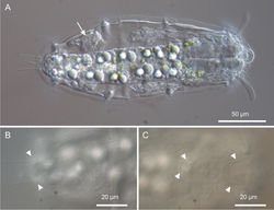
Variability and remarks. The largest adult was about 310 μm in total length (terminal spines excluded) and was carrying a very big egg inside (73 × 107 μm, Figure 3A). Remarkably, the egg shell was ornated with spikes (Figure 3B); this is quite surprising because in freshwater Gastrotricha the shell ornamentation is believed to appear only after the egg has been laid, probably due to osmotic differences between the external vs internal milieu (Hummon 1984[2]). The smallest animal was about 183 μm in total length and had no recognizable female gametes inside. Oocytes became appreciable as such in a 230 μm long specimen (Figure 4A).
Taxonomic affinities
The general body appearance, the presence of club-shaped cephalic tentacles and the planktonic lifestyle that characterise Kijanebalola devestiva sp. n. suggest that the closest affiliation of the South African specimens lies with the Neogosseidae. Notwithstanding this, autoapomorphic traits of the new species make somewhat difficult its affiliation to any of the two currently recognized genera, Neogossea and Kijanebalola, based on their current diagnosis (see Kisielewski 1991[3]). For instance, the size of the South African worms exceed by far that of any other possible keen (max length 310 μm vs 200 μm in Neogossea spp., vs 210 in Kijanebalola spp.) and the body covering, made for most part of smooth cuticle, is a characteristic so far unknown among species of Neogossea or Kijanebalola. The presence in the new species of a single cephalic plate (i.e., the cephalion), combined with the structure of its mouth and pharynx, complicate further its taxonomic affiliation at generic level. However, in our opinion, some of the differences between the anatomical traits of the iSimangaliso gastrotrichs and those highlighted in the diagnosis of Neogossea or Kijanebalola no longer hold. Regarding Neogossea, for instance, the diagnosis: 1) is ambiguous about the presence of cephalic plates (i.e., cephalion and hypostomion reported as undetected); and 2) includes the mouth as consisting of two-segmented units (mouth units = internal cuticular ridges). A re-evaluation of these characteristics, especially in light of the new information gained by Kieneke and Riemann (2007)[4] on German specimens of Neogossea (reported as Neogossea voigti (Daday, 1905) allows for the amendment of the generic diagnosis of Neogossea in both these aspects. More specifically, the specimens studied by Kieneke and Riemann (2007)[4] under differential interference contrast and scanning electron microscopy show: 1) the hypostomion and 2) the mouth units as unsegmented. Considering that in Neogosseidae the presence of a cephalion may be elusive (as reported above for the new species) we conclude that the presence/absence of the cephalic plates and the structure of the mouth and pharynx cannot be considered diagnostic characters at genus level (i.e., useful to distinguishing Neogossea from Kijanebala and vice versa); rather, peculiarities in these traits and others such as the presence/absence of cuticular reinforcements in the anterior pharyngeal bulb, may help differentiate among species (i.e., they are species specific).
Another potential difference between the two genera i.e., the presumed ability of Kijanebalola species to partially retract the head inside the body, can also be dismissed. Our observations with the new species demonstrate that retraction of the head is actually an artefact due to fixation (see above). Consequently, none of the above reported traits may be used to allocate our specimens to either of the two genera.
In summary, only a single character is apparently left to differentiate Neogossea and Kijanebalola; it pertains to the structure of the posterior part of the trunk, which appears truncate with a pair of postero-lateral projections, each provided with a tuft of relatively long spines in Neogossea, but rounded-off with a median group of spines in Kijanebalola. As the difference in this character between species of the two genera is consistent, it is reasonable to use it as diagnostic feature and as a solid synapomorphy for each genus.
Consequently, based on the round shape of the posterior trunk region of the iSimangaliso specimens, we propose that their closest affiliation is to the genus Kijanebalola. Therefore, the large size, the cuticular ornamentation reduced to an epaulet of scales on the ventral side of the trunk ending, and the number and size of the terminal spines are considered autoapomorphic characters that can easily distinguish Kijanebalola devestiva sp. n. from Kijanebalola dubia and Kijanebalola canina. Emended diagnoses are reported below.
Original Description
- Todaro, M; Perissinotto, R; Bownes, S; 2013: Neogosseidae (Gastrotricha, Chaetonotida) from the iSimangaliso Wetland Park, KwaZulu-Natal, South Africa ZooKeys, 315: 77-94. doi
Other References
- ↑ Kieneke A, Ahlrichs W, Arbizu P, Bartolomaeus T (2008a) Ultrastructure of protonephridia in Xenotrichula carolinensis syltensis and Chaetonotus maximus (Gastrotricha: Chaetonotida): comparative evaluation of the gastrotrich excretory organs. Zoomorphology 127: 1-20.
- ↑ Hummon M (1984) Reproduction and sexual development in a freshwater gastrotrich. 1. Oogenesis of parthenogenic eggs (Gastrotricha). Zoomorphologie 104: 33-41. doi: 10.1007/BF00312169
- ↑ Kisielewski J (1991) Inland-water Gastrotricha from Brazil. Annales Zoologici (Warsaw) 43 Suplement 2: 1–168.
- ↑ 4.0 4.1 Kieneke A, Riemann O (2007) Eine interessante Teilgruppe der Süßwassergastrotrichen (Bauchharlinge): Neogosseidae und Dasydytidae. Microcosmos 96: 139-146.
Images
|


