Ilyphagus wyvillei
| Notice: | This page is derived from the original publication listed below, whose author(s) should always be credited. Further contributors may edit and improve the content of this page and, consequently, need to be credited as well (see page history). Any assessment of factual correctness requires a careful review of the original article as well as of subsequent contributions.
If you are uncertain whether your planned contribution is correct or not, we suggest that you use the associated discussion page instead of editing the page directly. This page should be cited as follows (rationale):
Citation formats to copy and paste
BibTeX: @article{Salazar-Vallejo2012ZooKeys190, RIS/ Endnote: TY - JOUR Wikipedia/ Citizendium: <ref name="Salazar-Vallejo2012ZooKeys190">{{Citation See also the citation download page at the journal. |
Ordo: Terebellida
Familia: Flabelligeridae
Genus: Ilyphagus
Name
Ilyphagus wyvillei (McIntosh, 1885) – Wikispecies link – Pensoft Profile
- Trophonia wyvillei McIntosh, 1885:366–370, pl. 44, fig. 6, pl. 23A, figs 11–14, pl. 36A, figs 5–7, pl. 37A, fig. 1.
- Ilyphagus wyvillei:Hartman 1966[1]:41–43, pl. 12, figs 7, 8 (n. comb.); Levenstein 1975[2]:133; Detinova 1993[3]:100–101.
- Brada gravieri McIntosh, 1922:7–8, pl. 1, figs 4–6, pl. 3, fig. 1; Hartman 1966[1]:33, pl. 9, figs 1, 2; Hartman 1978[4]:173.
Type material
Southeastern Pacific Ocean. Holotype (NHML-85.12.1.261), R/V Challenger Expedition, Stat. 157 (53°55'S, 108°35'E), dredged, 1950 fathoms (3568.5 m), diatom ooze, 3 Mar. 1874.
Additional material
Antarctic Ocean. Several specimens (SIORAS-unnumb.), R/V Akademik Kurchatov, Stat. 914 (56°21'S, 50°48'W), 5650–6070 m, 14 Dec. 1971 (best specimen 49 mm long, 10 mm wide, cephalic cage 24 mm long (chaetiger 2 notochaetae 18 mm long), 22 chaetigers; 11 notochaetae in chaetiger 1; two anterior fragments dissected).
Description
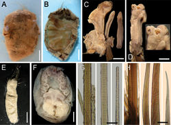
Holotype pale brown (Fig. 5A), completely dissected mid-ventrally (Fig. 5B), internal organs and most anterior end appendages previously removed (now lost). Body sausage-shaped, anteriorly truncate, medially widened, posteriorly rounded (confirmed in non-type specimen, Fig. 5E); 63 mm long, about 30 mm wide, cephalic cage 27 mm long, 19 chaetigers. Body surface papillated; papillae abundant, cylindrical, very long, sediment particles along papillae, more abundant basally.
Anterior end dissected, most appendages now lost. Cephalic hood short, margin smooth. Prostomium flat, without eyes. No caruncle. Palps very large; one remains attached to anterior end fragment (Fig. 5C), with a distal parasite (Fig. 5D); other palp loose in container , longer than branchiae (longest remaining detached branchia 5 mm long), expanded, with a median furrow; palp lobes reduced. Branchiae cirriform, distally colorless, sessile on branchial plate, arranged in single row, in horse-shoe pattern (Fig. 5F), with 16 filaments (perhaps other four, much smaller, filaments distally, would make a secondary distal row).
Cephalic cage chaetae as long as half body length, or about as long as body width. Chaetigers 1–2 involved in the cephalic cage, chaetiger 1 with 8–9 notochaetae in a single transverse row and 11–12 neurochaetae arranged in a C-pattern, opening to posterior region; chaetiger 2 with 5–6 noto- and 9–10 neurochaetae.
Anterior dorsal margin of first chaetiger truncate, papillated; anterior chaetigers without especially long papillae. Chaetigers 1–3 becoming progressively longer. Chaetal transition from cephalic cage to body chaetae abrupt; aristate neurospines from chaetiger 3. Gonopodial lobes in chaetiger 5, short, dark digitate, mostly covered by papillae.
Parapodia poorly developed, chaetae emerge from the body wall. Parapodia lateral, median neuropodia ventrolateral. Noto- and neuropodia close to each other, without especially longer papillae, some slightly thicker papillae bordering chaetae.
Median notochaetae arranged in short transverse rows, most notochaetae broken, length relationships with body width unknown, 1–3 per ramus; all multiarticulated capillaries, articles short basally, slightly longer medially, long subdistally (tips unknown, Fig. 5G). Neurochaetae multiarticulated capillaries in chaetigers 1–2; aristate neurospines from chaetiger 3, arranged in transverse rows, 7–8 per bundle. Each neurospine with very short articles basal- and medially (Fig. 5H); distally hyaline, smooth.
Posterior end rounded; pygidium with anus ventro-terminal, without anal cirri.
Remarks
Ilyphagus wyvillei (McIntosh, 1885) resembles Ilyphagus bythincola because they both have globose bodies with distally smooth aristate neurospines. They differ because Ilyphagus wyvillei has comparatively shorter cephalic cage chaetae than Ilyphagus bythincola, and because in Ilyphagus wyvillei there are only 16 branchiae, whereas in Ilyphagus bythincola there are about 40. On the other hand, Ilyphagus wyvillei resembles Ilyphagus coronatus Monro, 1939, because in their first chaetiger, neurochaetae are arranged in a C-pattern, opening to the posterior region, and by having distally smooth neurospines. However, they differ because Ilyphagus wyvillei has fewer chaetigers (19–22 vs 23–25) and a more globose body but these differences might be modified after more specimens are studied. Two other differences are probably more relevant and must be emphasized:the relative number of neurochaetae in the first chaetiger (11–12 in Ilyphagus wyvillei, 8 in Ilyphagus coronatus) , and the start of the aristate neurospines (chaetiger 2 in Ilyphagus wyvillei, chaetiger 3 in Ilyphagus coronatus).
The presence of parasitic copepods in the branchial bases of Ilyphagus wyvillei cannot be confirmed due to the state of the anterior end; however, one portion of a parasite is visible at one of the palps tip, and there is another deep scar in the same palp. McIntosh might have confused the attachment site, because he dissected the anterior end and branchial scars could be confused with these parasite attachment sites.
Brada gravieri McIntosh, 1922 might belong to the same species. There is no type material available; it is probably lost. However, the original illustrations and description noticed the lack of the cephalic cage chaetae, whereas the neurochaetae (pp 7–8) were described as translucent, smooth, devoid of transversal marks. The distal part of neurochaetae is often smooth, hyaline, but the rest of the chaetae have anchylosed articles or transverse markings throughout it. They were collected from relatively close localities but fresh material needs to be examined to clarify this .
Distribution
Originally described from the Antarctic Ocean, it has been found in abyssal depths off Western South America (Levenstein, 1975). The Bering Sea records by Levenstein (1961a[5]:160, 1966[6]:46), cannot be confirmed because the specimens were not found.
Taxon Treatment
- Salazar-Vallejo, S; 2012: Revision of Ilyphagus Chamberlin, 1919 (Polychaeta, Flabelligeridae) ZooKeys, 190: 1-19. doi
Other References
- ↑ 1.0 1.1 Hartman O (1966) Polychaeta Myzostomidae and Sedentaria of Antarctica. Antarctic Research Series 7: 1-158. doi: 10.1029/AR007
- ↑ Levenstein R (1975) Mnogotschetinkovye chervi (Polychaeta) zhelobov Atlantitcheskogo sektora Antarktiki. Akademiya Nauk SSSR, Trudy Instituta Okeanologii 103: 119-142.
- ↑ Detinova N (1993) Mnogoshchetinkovye chervi Orkneiskogo zheloba. Trudy Instituta Okeanologiya P.P. Shirshova, Rossiya Akademi Nauk 127: 97-106.
- ↑ Hartman O (1978) Polychaeta from the Weddell Sea Quadrant, Antarctica. Antarctic Research Series 26: 125-222. doi: 10.1029/AR026p0125
- ↑ Levenstein R (1961a) Mnogotschetinkovye tchervi glubokovodnoi tchasti Beringova morya. Akademiya Nauk SSSR, Trudy Instituta Okeanology 46: 147-178.
- ↑ Levenstein R (1966) Mnogotshchetinkov’e chervi (Polychaeta) zapadnoi tasti Beringova morya. Akademiya Nauk SSSR, Trudy Instituta Okeanologii 81: 3-131.
Images
|
