Homidia jordanai
| Notice: | This page is derived from the original publication listed below, whose author(s) should always be credited. Further contributors may edit and improve the content of this page and, consequently, need to be credited as well (see page history). Any assessment of factual correctness requires a careful review of the original article as well as of subsequent contributions.
If you are uncertain whether your planned contribution is correct or not, we suggest that you use the associated discussion page instead of editing the page directly. This page should be cited as follows (rationale):
Citation formats to copy and paste
BibTeX: @article{Pan2011ZooKeys152, RIS/ Endnote: TY - JOUR Wikipedia/ Citizendium: <ref name="Pan2011ZooKeys152">{{Citation See also the citation download page at the journal. |
Ordo: Entomobryomorpha
Familia: Entomobryidae
Genus: Homidia
Name
Homidia jordanai Pan & Shi & Zhang, 2011 sp. n. – Wikispecies link – ZooBank link – Pensoft Profile
Holotype
♂ on slide, Shaoxin City, Zhuji Country, Dongbaihu, Zhejiang Province, CHINA, 29°34.48'N, 120°24.32'E, 3.X.2009, collection number S4014, collected by Zhi-Xiang Pan and Chen-Chong Si, deposited in Taizhou University.
Paratypes
2 ♂, 11 ♀ and 5 larvae on slide, numerous in alcohol, same data as holotype. 5 paratypes (1 ♂, 1 ♀ on slide, 1 larva and 2 adults in alcohol) deposited in School of Life Sciences, Nanjing University and others in Taizhou University, China.
Etymology
Named after the famous Spanish entomologist Jordana Rafael (University of Navarra).
Description
Adult. Size. Maximum body length up to 2.3 mm.
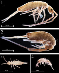
Habitus. Ground colour pale yellow in alcohol. Body dorsally without pigment. Coxa and trochanter of all legs with weak blue pigment. Eye patches dark. Antennae gradually darker from Ant. III to Ant. IV (Figs 1, 2).
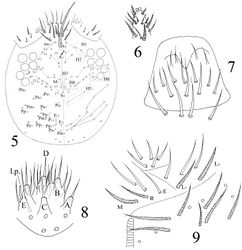
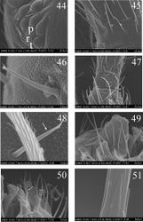
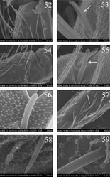
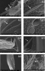
Head. Ommatidia 8+8, G and H smaller than others, and sometimes invisible under LM, interocular setae as p, r, t (Figs 5, 44). Antenna 3.9–4.6 times as long as cephalic diagonal, subequal to body in length, antennal segment ratio as I : II : III : IV = 1 : 1.3–1.5 : 1.3–1.4 : 2.4–3.1. Basal Ant. I with 2 dorsal and 4 ventral spiny setae (Figs 5, 45, 46). Basal Ant. II with 5 smooth setae (Figs 47, 48); distal Ant. II with 4 s (2–3 longer, 1–2 shorter) (Figs 52, 53). Ant. III organ with 2 rod-like s and 3 small s (Figs 54–56); those s also with obvious ridges on surface under SEM. Distal Ant. IV with several types of s (Figs 57–62); apical bulb bilobed (Fig. 6). Dorsal cephalic chaetotaxy with 4An and 7S mac. Clypeus with many ciliate setae (Fig. 5). Labral papillae absent. Prelabral and labral setae as 4/5, 5, 4, all smooth, labium intrusion U-shaped (Fig. 7). Maxillary outer lobe with 1 apical seta, 1 subapical seta and 3 sublobal hairs on sublobal plate; subapical seta slightly longer than apical one (Fig. 49). Labial palp with five papillae A–E, with 0, 5, 0, 4, 4 guard setae, respectively, and 5 smooth proximal smooth setae; lateral process differentiated with blunt tip reaching apex of papilla E (Figs 8, 50, 51). Hyaline plate with 1 main (H) and 2 accessorial (h1, h2) setae. Setal formula of labial base as MREL1L2, seta E smooth, others ciliate (Fig. 9).
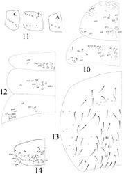
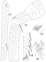
Thorax. Complete s-chaetae of dorsal body as 32/223(>47)3 (examined specimens mostly with 47 s-chaetae on Abd. IV, but some lost during preparation), ms as 10/10100. Th. II with 3 medio-medial (m1, m2, m2i), 3 medio-sublateral (m4, m4i, m4p), 17–18 posterior mac and 3 s-chaetae (ms antero-internal to s); seta p1i2 rarely present, seta p6 as mic. Th. III with 29–32 mac and 2 s-chaetae; setae p1i2 and p4 absent (Fig. 10). Numerous setae on hind leg (Figs 15–19); pseudopores on coxa shown in Fig. 63, but their number unclearly seen. Coxal macrochaetal formula as 3/4+1, 2+1/4+2 (Fig. 11). Trochanteral organ with 36–64 smooth spiny setae (Fig. 16). Inner differentiated tibiotarsal setae slightly ciliate, most distal smooth seta present on hind leg (Figs 18, 19, 65). Tenent hairclavate and subequal to inner edge of unguis in length (Figs 19, 66). Unguis with 4 inner, 2lateral and 1 outer teeth, all tiny. Unguiculus lanceolate with outer edge slightly serrate (Fig. 65). Pretarsus with 1 pair of small spines (Figs 19, 67).
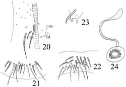
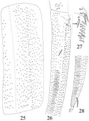
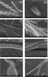
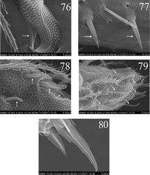
Abdomen. Abd. I with 9 (a2, a3, m2–4, m2i, m4i, m4p, a5) mac and 2 s-chaetae (ms anterio-external to s) (Figs 68, 69). Abd. II with 6 (a2, a3, m3, m3e, m3ea, m3ep) inner, 1 (m5) lateral mac, and 2 s-chaetae. Abd. III with 1 (m3) inner and 4 (am6, pm6, m7a, p6) lateral mac, and 3 s-chaetae (Fig. 12). Abd. IV with more than 47 s-chaetae (2 of normal length and others elongated), 6–9 mac on anterior part and irregularly arranged in a transverse row; posterior part with 2 (3) (B5 and A6, A6 rarely as mic, B4 sometimes present) mac and 1 (B6) mic (Fig. 13). Abd. V with 3 s-chaetae; m3a absent, a5i sometimes absent (Fig. 14). Anterior face of ventral tube with many ciliate setae, including 4+4 mac, line connecting proximal (Pr) and external-distal (Ed) (Chnd and Li 1997) mac oblique to median furrow(Fig. 20); posterior face with 5 or 6 (median with 1 or 2 small) smooth and numerous ciliate setae (Fig. 21); lateral flap with 6–7 smooth and 10–22 ciliate setae (Figs 22, 74, 75). Furcula shown in Figs 25–28. Manubrial plaque with 3 pseudopores, 2 inner and 5–6 outer ciliate setae (Fig. 26). Dens with 20–40 spines (Figs 26, 27, 77); basal sete (Szeptycki 1973[1]) bs1 and bs2 spiny and multilaterally ciliate, bs1 shorter than bs2; proximal-inner seta (pi) spiny, shorter and thicker than bs1 and bs2 (Figs 26, 27). Mucro bidentate with subapical tooth obviously larger than apical one; basal spine short, with tip only reaching apex of subapical tooth (Figs 28, 76). Tenaculum with 4+4 teeth and 1 large, multilaterally ciliate basal seta (Fig. 23). Genital plate papillate (Fig. 24).
The first instar larva
Size. Body length up to 0.7 mm.
Habitus. Ground colour pale white in alcohol. Eye patches dark. Distal antennae slightly pigmented (Figs 3, 4).
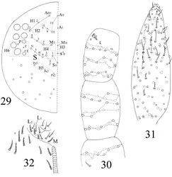
Head. Antenna 1.3–1.8 times as long as cephalic diagonal, antennal segment ratio as I : II : III : IV = 1 : 1.4–2.2 : 1.5–2.5 : 3.3–4.3. Ant. I with 11 ciliate and 1 basal spiny setae. Ant. II with 25 ciliate setae. Ant. III with 38 ciliate setae and 5 s-chaetae of Ant. III organ (Figs 30, 78). Ant. IV with numerous ciliate setae and some s-chaetae (Figs 31, 79). Dorsal cephalic chaetotaxy as 3An and 5S mac (Fig. 29). Labium with 3 smooth proximal setae. Setal formula of labial base as MEL1L2, seta E smooth, all others ciliate (Fig. 32). Ommatidia, Ant. IV apical bulb, interocular setae, labral papillae, labrum, maxillary outer lobe, labial palp, hyaline plate same as adults.
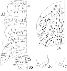
Chaetotaxy. Complete s-chaetae of body as 32/223(50–53)3, ms as 10/10100. Th. II with 13 (a1–6, m1, m4, m6, p1–3, p5) mac, 6 (a7, m2, m5, m7, p4, p6) mic and 3 s-chaetae (ms anterior to s), m1 rarely as mic. Th. III with 9 (a2–6, m6, p1–3) mac, 9 (a1, a7, m1, m4, m5, m7, p4, p6) mic and 2 s-chaetae. Abd. I with 3 (m2–4) mac, 9 (a1–3, a5, a6, m5, m6, p5, p6) mic and 2 s-chaetae (ms antero-external to s). Abd. II with 2 (m3, m5) mac, 13 (a1–3, a6, a7, m4, m6, m7, p4, p7, el) mic, 1 additional mic on lateral and 2 s-chaetae. Abd. III with 1 (m3) mac central, 13 (a1–3, a6, a7, m4, am6, pm6, m7, p4) mic, 3 s-chaetae and 5 lateral additional mic (Fig. 33). Abd. IV with 2 (B5, E3) mac, 27 (A1–4, A6, B1–4, B6, C1–4, T1, T3, T5, D1–3, E1, E4, F1–3) mic and 50–53 s-chaetae (48–51 elongated and 2 of normal length); setae A5 and E2 absent (Fig. 34). Abd. V with total 14 setae and 3 s-chaetae. Abd. VI with 21+21 setae; 3 on middle line (Fig. 35).
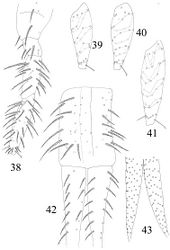
Leg. Coxa I–III with 2, 3, 5 ciliate setae. Trochanter I–III with 4, 5, 4 ciliate and 2, 1, 0 smooth setae, 1 spine on trochanter III. Femur I–III with 10, 16, 14 ciliate and 3, 1, 3 smooth setae. Tita I–III with 38, 41, 46 ciliate setae and 1 tenent hair respectively, 1 supraempodial seta on Tita III (Figs 38–41). Unguis with 4 minute inner and 2lateral teeth. Unguiculus lanceolate with outer edge slightly serrate (Fig. 80).
Ventral tube. Anterior face without seta; posterior face with 2 apical smooth setae; lateral flap with 2 smooth setae (Fig. 36).
Furcula. Manubrum with 24+24 ciliate setae. Manubrial plaque not seen. Dorsal dens with 14–15 (8 in outer 6–7 in inner row) setae, ventral side with about 55 ciliate setae, inner of dens without dental spine, basal setae (bs1 and bs2) absent (Figs 42, 43). Mucro bidentate with subapical tooth obviously larger than apical one; basal spine absent. Tenaculum with 4+4 teeth but without setae on corpus (Fig. 37).
Ecology
In the leaf litter of Cunninghamia lanceolata (Lambert) and Dicranopteris dichotoma (Thunberg).
Remarks
The new species is characterized by weak pigment on dorsal body, long antennae subequal to body in length, labial basal seta L1 ciliate, absence of setae m2i2 on Th. II, a1 on Abd. I, a2 on Abd. III, A4–5 on Abd. IV, and dental basal seta pi spiny and shorter than bs. It is similar to Homidia unichaeta Pan, Shi & Zhang, 2010 and Homidia tibetensis Chen & Zhong, 1998 in colour pattern, cephalic chaetotaxy, labrum, coxal formula, chaetotaxy of Abd. II and claw. However, it can be distinguished from them by the length of antennae, labial setal formula, chaetotaxy on Th. II, Abd. I, Abd. III–IV and seta pi on basal dens. Additional differences are listed in Table 1.
Differences between the first instar larvae and adults
Some characters are principally same in the first instar larvae and adults: ommatidia, interocular setae, Ant. III organ, apical bulb on Ant. IV, labrum and labral papillae, labial palp, maxillary outer lobe, claw, bothriotricha and s-chaetoxic pattern on terga.
Characters that develop after the first instar: s on distal part of Ant. II, smooth setae on base of Ant. II, labial seta R, smooth spiny setae on trochanteral organ, mac on anterior face of ventral tube, seta on corpus of tenaculum, pseudopores on manubral plaque, basal spine on mucro and genital plate.
Chaetotaxy become more complicated during postembryonal development, detailed differences between the first instar larvae and adults (apart from chaetotaxy of body tergites) are listed in Table 2. Tergal chaetotaxy of adults becomes much more complicated than that of primary chaetotaxy. In addition to numerous secondary common mic and mac on terga, some primary mic are transformed into mac: m2, m5 and p4 on Th. II, a1, m5, p5 and p6 on Th. III, a2, a3 and a5 on Abd. I, a2 and a3 on Abd. II, am6 and p6 on Abd. III, A6 and B4 on Abd. IV (homology of lateral setae difficult to determine).
| Characters | H. j | H. u | H. t |
| Pigment on dorsal tergite | - | - | slight |
| Antenna length as long as cephalic diagonal | 3.9–4.6 | 1.5–2.5 | about 3.5 times |
| Seta L1 on labial base | ciliate | smooth | smooth |
| Lateral prosess of labial papilla E | reach apex | not reach | ? |
| Seta m2i2 on Th. II | - | + | - |
| Seta a1 on Abd. I | - | + | + |
| Seta a2 on Abd. III | - | + | + |
| Mac on posterior Abd. IV | 2 (rarely 3) | 1 | 2 |
| “Eyebrow” setae on anterior Abd. IV | 6–9 | 3–8 (usually 5–7) | 10–12 |
| Dental spines | 20–40 | 19–23 | 39–54 |
| Comparison of dental basal seta in length | bs > pi | pi > bs | bs > pi |
| Type locality (China) | Zhejiang | Zhejiang | Tibet |
|+ Table 2. Differences between the first instar larvae and adults (apart from chaetotaxy of body tergites). |- | Characters || First instar larvae || Adults |- | Ground colour || pale white || pale yellow |- | Cephalic chaetotaxy || 3An, 5S || 4An, 7S |- | Spiny setae on basal Ant. I || 1 || 2 dorsal, 4 ventral |- | S on distal Ant. II || - || 4 |- | Proximal setae of labium || 3 || 5 |- | Seta R on labial base || - || + |- | Coxal macrochaetal formula || 2/3/5 || 3/4+1, 3/4+2 |- | Setae on trochanteral organ of hind leg || 1 || 36–64 |- | Inner tibiotarsal setae || strongly ciliate || thicker and slightly serrate |- | Setae on anterior face of ventral tube || - || 4+4 mac and numerous mic |- | Setae on lateral flap of ventral tube || 2 smooth || 6–7 smooth and 10–24 ciliate |- | Setae on posterior face of ventral tube || 2 smooth || 5 or 6 smooth and numerous ciliate |- | Seta on tenaculum || - || 1 |- | “Eyebrow” setae on Abd. IV || - || 6–9 |- | Setae on manubrium || 24+24 || more than 24+24 |- | Pseudopores on manubrial plaque || - || 3 |- | Ciliate setae on manubrial plaque || - || 7–8 |- | Basal setae (bs1 and bs2) on dens || - || + |- | The shape of proximal-inner seta (pi) || normal || spiny |- | Dental spines || - || 20–40 |- | Basal spine of mucro || - || + |- | Genital plate || - || papillate |} {| class="wikitable" ; style="width: 100%" |+ Table 3. Morphological differences of the first instar larvae among six Entomobyridae species. |- | tergite || seta || H. j || O. f || H. n || E. m || P. a || S. d |- | Th. II || m1 || mac || mac || mic || mac || mac || mac |- | || m2 || mic || mac || mic || mic || scale || mac |- | || p5 || mac || mac || mic || mic || mac || mac |- | Th. III || a2 || mac || mic || mic || mic || mic || mac |- | || a3 || mac || mic || mic || mic || mic || mic |- | || a4 || mac || mac || mic || mic || mac || mac |- | || m1 || mic || mic || mic || mic || - || mic |- | || m2 || - || - || - || - || mac || - |- | Abd. I || m2 || mac || mac || mac || mic || mac || mac |- | || m4 || mac || mac || mic || mac || mac || mac |- | || m6 || mic || mic || mic || mic || mic || mic |- | Abd. II || a2 || mic || mac || mic || mic || mac || mac |- | || m5 || mac || mac || mic || mac || mac || mac |- | Abd. III || pm6 || mac || mac || mac || mac || mac || mac |- | || p6 || mic || mic || mic || mac || mic || mac |- | Abd. IV || A4 || mic || - || - || mic || - || - |- | || A5 || - || - || - || - || mic || mic |- | || B4 || mic || - || - || mic || mic || mac |- | || B5 || mac || mic || mic || mac || mac || mac |- | || B6 || mic || mic || - || mic || mic || mic |- | || E2 || - || mac || mic || - || mic || mic |- | || E3 || mac || - || - || mac || mic || mac |}
Original Description
- Pan, Z; Shi, S; Zhang, F; 2011: New species of Homidia (Collembola, Entomobryidae) from eastern China with description of the first instar larvae ZooKeys, 152: 21-42. doi
Other References
- ↑ Szeptycki A (1973) North Korean Collembola. I. The genus Homidia Börner, 1906 (Entomobryidae). Acta Zoologica Cracoviensia 31: 23-39.
Images
|
