Hexabranchus sanguineus
| Notice: | This page is derived from the original publication listed below, whose author(s) should always be credited. Further contributors may edit and improve the content of this page and, consequently, need to be credited as well (see page history). Any assessment of factual correctness requires a careful review of the original article as well as of subsequent contributions.
If you are uncertain whether your planned contribution is correct or not, we suggest that you use the associated discussion page instead of editing the page directly. This page should be cited as follows (rationale):
Citation formats to copy and paste
BibTeX: @article{Yonow2012ZooKeys197, RIS/ Endnote: TY - JOUR Wikipedia/ Citizendium: <ref name="Yonow2012ZooKeys197">{{Citation See also the citation download page at the journal. |
Familia: Hexabranchidae
Genus: Hexabranchus
Name
Hexabranchus sanguineus (Rüppell & Leuckart, 1828) – Wikispecies link – Pensoft Profile
- Hexabranchus sanguineus. – Yonow & Hayward 1991: 15, fig. 3D (Mauritius); Valdés 2002[1]: 291, figs. 1A, C, 2-4 (South Africa, Mozambique Channel, Madagascar, Philippines, Hawaii) incl. extensive synonymy.
- Hexabranchus flammulatus (Quoy & Gaimard). – Narayanan 1968[2]: 378, fig. 2 (India).
- Hexabranchus marginatus (Quoy & Gaimard). – Edmunds 1971[3]: 340 (Tanzania); Richmond 2011[4]: 280 (East Africa).
- Hexabranchus sp. – Rudman 1986: 347, figs. 1, 19, 20 (Tanzania and Christmas Island).
Material
Socotra: 55 mm pres. length (WPU Rostock, MAR 85, RC-N57), Hadibo, late 1980s, leg. W Wranik. – Yemen: 85 × 60 mm pres., distorted and flattened (WPU Rostock, MAR 85, RC-N16), Al Mukalla, late 1980s, leg. W Wranik. – Kenya: two specimens 60 and 95 mm in length, pres., Vipingo, 25 m N of Mombasa, ELW in rock pools on exposed reef, 23 September 1984, leg. J Hognerud. – Tanzania: photographs of one individual, M’Nazi Bay, Msimbati, near Mtwara, May 1994, IM Horsfall. – Maldives: one juvenile specimen 13 mm (MDV/AB/96/17), Yacht Tila, South Malé Atoll, 25 m depth, 09 May 1996, leg. RC Anderson & SG Buttress; photos of 15 mm juvenile individual, 1986-1994, J Hinterkircher. – La Réunion, Mauritius, Mayotte: photographs of numerous individuals, including juveniles similar to the Maldives specimen examined here (http://seaslugs.free.fr/nudibranche/a_intro.htm) and slides of 27 individuals varying from yellow to mottled pink, M Parmantier. – Seychelles: photos of several individuals including juvenile and sub-adult, Lilôt, NW Mahé, 1988-1989, P Kemp. – Sri Lanka: photographs of one large individual, Unawatuna, S of Galle, on sediment-covered rock, 27 December 2011, S Kahlbrock.
Description
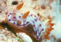
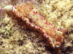
The large specimens and individuals were of the typical Indo-West Pacific colour pattern, blotchy red and cream, especially along the margins. The 13 mm juvenile specimen from the Maldives illustrated here (Plate 26) is well relaxed, and the six gills can be seen to insert into separate openings. The oral tentacles are large rounded lobes, clearly the precursors to the lappets of the adult: these tentacles are huge in comparison to those of a chromodorid (or dorid) of similar size. The rhinophores have 13 lamellae; note that the white edges present in the adults have not yet developed. Another 15 mm juvenile individual was identical in colour pattern, while a slightly larger animal from the Seychelles demonstrated the developing adult colour pattern already had white edges to the 17+ rhinophore lamellae (Plate 27).
The radular formulae of the larger specimens are tabulated below: the sizes listed are of the same dimension of a large lateral tooth – from the tip of the cusp to the flange where the cusp meets the base. There is no relationship between the radular formula and maximum tooth size, nor are they correlated with preserved animal size. A giant Hong Kong specimen is included for comparison, and has the largest teeth but not the largest radula. It had a bubbly texture and was pinkish yellow in life (M Collard pers. comm.; specimen, radula, notes and photographs lodged in the Natural History Museum, London: NHMUK acc. no. 2337 with the “Red Sea Giant”).
Socotra 55 mm pres. 41 × 64.0.64 500 μm
Yemen 85 mm pres. 53 × 93-83.0.83-93 550 μm
Kenya 90 mm pres. 47 × 77.0.77 700 μm
Kenya 95 mm pres. 46 × 79.0.79 450 μm
Hong Kong 190 mm pres. 51 × 67.0.67 800 μm
Remarks
Yonow (2001)[5] suggested that the uniformly red species in the Red Sea should be assigned to Hexabranchus sanguineus (Rüppell & Leuckart), and that it was distinct from the widespread Indo-Pacific Hexabranchus marginatus (Quoy & Gaimard). The study mentioned in that paper was never published, since the following year a paper analyzing the same problem was published by Valdés (2002)[1]: Valdés examined material from the western Indian Ocean and the Pacific, but no Red Sea specimens were included in his analysis, nor were any ‘giants’ but, despite this, he concluded that all Indo-Pacific species were the same, and that only the Caribbean species Hexabranchus morsomus Ev. Marcus & Er. Marcus was distinct.
Taxon Treatment
- Yonow, N; 2012: Opisthobranchs from the western Indian Ocean, with descriptions of two new species and ten new records (Mollusca, Gastropoda) ZooKeys, 197: 1-130. doi
Other References
- ↑ 1.0 1.1 Valdés A (2002) How many species of Hexabranchus (Opisthobranchia: Dorididae) are there? Molluscan Research 22: 289–301. doi: 10.1071/MR02012
- ↑ Narayanan K (1968) On three opisthobranchs from the south-west coast of India. Journal Marine Biological Association, India 10 (2): 377-380.
- ↑ Edmunds M (1971) Opisthobranchiate Mollusca from Tanzania (Suborder: Doridacea). (III). Journal Linnean Society (Zoology) 50: 339-396. doi: 10.1111/j.1096-3642.1971.tb00767.x
- ↑ Richmond M (2011) A Field Guide to the Seashores of Eastern Africa and the Western Indian Ocean Islands, 3rd edition. Sida/SAREC-UDSM, Sweden, 464 pp.
- ↑ Yonow N (2001) Results of the Rumphius Biohistorical Expedition to Ambon (1990). Part 11. Doridacea of the families Chromodorididae and Hexabranchidae (Mollusca, Gastropoda, Opisthobranchia, Nudibranchia), including additional Molukkan material. Zoologische Mededelingen, Leiden 75(1–15): 1-50.
Images
|

