Deuterophlebia pseudopoda
| Notice: | This page is derived from the original publication listed below, whose author(s) should always be credited. Further contributors may edit and improve the content of this page and, consequently, need to be credited as well (see page history). Any assessment of factual correctness requires a careful review of the original article as well as of subsequent contributions.
If you are uncertain whether your planned contribution is correct or not, we suggest that you use the associated discussion page instead of editing the page directly. This page should be cited as follows (rationale):
Citation formats to copy and paste
BibTeX: @article{Zheng2023DeutscheEntomologischeZeitschrift70, RIS/ Endnote: TY - JOUR Wikipedia/ Citizendium: <ref name="Zheng2023Deutsche Entomologische Zeitschrift70">{{Citation See also the citation download page at the journal. |
Ordo: Diptera
Familia: Deuterophlebiidae
Genus: Deuterophlebia
Name
Deuterophlebia pseudopoda Zheng & Zhou, 2023 sp. nov. – Wikispecies link – ZooBank link – Pensoft Profile
Description
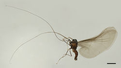
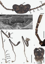
Male adults. Body length ca. 2.2–2.6 mm (n=5), uniformly brownish black (Fig. 1). Head brownish black, flattened, nearly trapezoidal, width ca. 0.50 mm, folded backward under thorax, hidden in dorsal view (Fig. 1). Head densely covered with microtrichia. Median clypeal lobe slightly convex, semicircle shaped, with around 20 sharp setae (Fig. 2A). Mouthparts in form of an invaginated tubule, oral region depressed (Fig. 2A, B). Edges of oral region (or mouth opening) ridged, convex medially on ventral ridge, forming a blunt mental tooth (Fig. 2A, B). Postgena and oral region with sparser microtrichia than other regions, a pair of tentorial pits present on each side of oral region (Fig. 2A, B). Compound eyes glabrous, width ca. 0.18 mm, distance between eyes ca. 0.30 mm (Fig. 2A). Antennae 8.5–10.0 mm (n=5) (Fig. 2A, C). Scape oval shaped, pedicel globular, both covered with microtrichia (Fig. 2A, C). Flagella four segmented, flagellomeres I–III slender cylindrical, each with a subapical tubercle on front margin, bearing 9–12, 6–9 and 4–6 digitiform setae respectively (Fig. 2C). Flagellomere IV flattened and elongated, broader than flagellomeres I–III, narrowed gradually, with curved hair-like setae on the anterior side of basal half, apical half generally glabrous but bearing 4–5 clusters of curved hair-like setae, apex slightly expanded with some curved hair-like setae (Fig. 2C). Antennal ratio: 4.0: 2.0: 5.0: 3.0: 3.0: 238.0 (Figs 1, 2C). Flagellomere IV about 14× combined length of five basal antennal articles or about 4× body length (Fig. 1).
Thorax uniformly brownish black, densely covered with microtrichia (Fig. 1). Pronotum almost hidden, mesonotum strongly expanded (Fig. 1). Wings ca. 4.0 mm, uniformly set with dark micro-tubercles, grayish translucent, cubital area greatly enlarged, costal margin slightly thickened (Fig. 1). Outer margin fringed with soft hair-like setae, denser and longer on cubital margin (Fig. 1). Veins radially arranged, pale and inconspicuous (Fig. 1). Halteres transparent, ca. 0.35 mm (Fig. 1).
Legs brownish black, slender, sharing similar chaetotaxy with four types of setae: (1) microtrichia, densely covered on all segments; (2) sharp macrotrichia, sparsely on dorsal margin of femora and tibiae; (3) long capitate setae, ventrally on tarsomeres I–IV of each leg, distal half of ventral edge of all tibiae, surrounding the top of fore- and midtibiae, and also densely arranged radially on each empodium; (4) digitiform setae, 1–3 pairs for each tarsomere (Fig. 2D–F). In all legs, coxae much broader than trochanters, coxae about twice the length of trochanters (Fig. 2D–F). In foreleg, femur: tibia: tarsus = 9.0: 14.0: 14.0; femur slightly flattened, tibia slender, cylindrical, and gradually broader apically; tarsomere I: II: III: IV: V = 6.4: 2.3: 1.4: 1.4: 1.0, tarsomeres I–IV cylindrical, tarsomere V conical; empodium shell-shaped, length subequal to tarsomere V; claw slender, tapered, shorter than empodium (Fig. 2D). Midleg shortest among all legs, similar to foreleg, femur: tibia: tarsus = 8.0: 10.5: 12.0, tarsomere I: II: III: IV: V = 6.0: 1.9: 1.1: 1.1: 1.0 (Fig. 2E). In hindleg, femur: tibia: tarsus of hindleg = 10.0: 13.5: 10.0, tarsomere I: II: III: IV: V = 3.5: 1.5: 1.2: 1.2: 1.0 (Fig. 2F).
Abdomen brownish black, densely covered with microtrichia, nine segmented, tapering posteriorly (Fig. 1). First two segments strongly fused with each other, paler and shorter than others (Fig. 1). Segment VIII in form of a short chitin ring (Fig. 1). Sternite IX almost glabrous, fused with dorsal plate, connected with gonocoxite (Fig. 2G). Gonocoxite with posterior projection which length subequal to gonostylus (Fig. 2G). Gonostylus subequal to the dorsal plate in length, flattened, oval-shaped, flexor surface with numerous curved sharp setae (Fig. 2G). Dorsal plate parallel-sided, posterior margin slightly depressed without cleft with some stout curved setae on margin (Fig. 2G). Aedeagus in form of a smooth tube, length subequal to gonostylus and dorsal plate (Fig. 2G).
Female adult. Body length ca. 2.0 mm (n = 1). Besides sexual differences, generally similar to the males except following features (Fig. 3A–D).
Head width ca. 0.36 mm (Fig. 3A). Median clypeal lobe strongly protruded medially with ca. 20 setae (Fig. 3A). Oral region located near anterior margin of head (Fig. 3A). Compound eyes more prominent than males (Fig. 3A). Antenna ca. 0.3 mm. Scape slender oval shaped, pedicel globular, both scape and pedicel covered with microtrichia and bearing several sharp setae (Fig. 3A). Flagellomere I slender cylindrical, flagellomeres II–III slender oval shaped, flagellomere IV dripping shaped, strongly narrowed basally (Fig. 3A). Each of flagellomeres I–III bearing ca. 5 digitiform setae apically, flagellomere IV with 4 sharp setae. Antennal ratio ca. 7.0: 3.0: 10.0: 4.0: 5.0: 4.0 (Fig. 3A). Legs sharing similar chaetotaxy and exhibiting three types of setae, chaetotaxy similar to males but without capitate setae (Fig. 3B–D). In foreleg and midleg, femur: tibia: tarsus = 1.0: 2.0: 1.3, tarsomere I: II: III: IV: V = 1.0: 0.8: 0.8: 0.8: 2.4. (Fig. 3B, C) In hindleg, femur: tibia: tarsus = 1.0: 1.7: 1.1, tarsomere I: II: III: IV: V = 1.0: 0.9: 0.9: 0.9: 2.7 (Fig. 3D). Claws of all legs similar, paired, stout and curved, with a blunt protrusion in the middle (Fig. 3B–D). Empodium in form of a long and hairy spine, subequal to the length of claw (Fig. 3B–D).
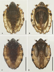
Male pupae. Pupae flattened oval shaped, length 2.3 mm (n = 2), width 1.6 mm. Dorsal integument dark brown, divided into 11 segments (Fig. 4A, B).
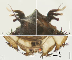
Prothorax fused with mesothorax, forming a conical segment with a median suture (Fig. 4A, B). Mesothoracic lateral margins each with a sharp spine and a gill (Figs 4A, B, 5A, B). Spines ca. 0.4 mm, slightly curved, dark brown, originated from a round base (Fig. 5A, B). Ventral gills light to dark brown, length subequal to the dorsal spines, hand-shaped and consisting of three filaments: posterior filament shorter, pointing backward; anterior two filaments similar in shape, twisted and light in color apically (Fig. 5A, B). Metathorax completely surrounded by mesothorax and first abdominal segment (Fig. 4A, B). Abdominal segments I–II with a pair of anterolateral projections, each projection pointing forward and bearing ca. 13 spines (Fig. 4A, B). Segments VI–VII with a pair of posterolateral projections, projections foot-shaped and each bearing ca. 8 spines (Fig. 5C). Segment VIII shield-shaped, surrounded by segments VII and IX (Fig. 4A, B). Adult structures visible on ventral side (Fig. 4A, B). Head present directly below mesothorax; antennal sheaths in form of a large elliptic ring, surrounding body 2.0 times (Fig. 4A, B). Leg sacs extended to posterior end of antennal ring, strongly expanded apically. Abdominal segments III–V with a pair of black adhesive discs (Fig. 4A, B).
Female pupae. Length ca. 2.2 mm (n = 2), width ca. 1.5 mm (Fig. 4C, D). Dorsal morphology similar to male except for smaller mesothorax (Fig. 4C, D). Gender can be identified through the absence of antennal ring, apex of female leg sheaths not expanded (Fig. 4C, D).
Material examined
Holotype: male adult, China: Yunnan Province, Gongshan County, Dulongjiang Township, Dulongjiang River, 27°50'14.16"N, 98°19'54.2"E, 1470 m a.s.l., 4.II.2023, Xuhongyi Zheng leg. Paratypes: 6 male adults, 1 female adult, 2 male pupae, 2 female pupae, same locality and data as holotype.
Diagnosis
Male adults of Deuterophlebia pseudopoda sp. nov. can be identified by their terminalia: gonostylus short, length of gonostylus subequal to the gonocoxite and dorsal plate; posterior margin of dorsal plate slightly depressed but without a median cleft (Fig. 2G). Such a terminalia differs from the 19 named Deuterophlebia species (Courtney 1990[1], 1994[2]; Zheng et al. 2022[3]). The shape of their heads is also distinct among known species: median clypeal lobe slightly convex, inner side of compound eye without a protruded corner (Fig. 2A) (Courtney 1990[1], 1994[2]; Zheng et al. 2022[3]).
Female adults of D. pseudopoda sp. nov. can be recognized through a combination of the pronounced median clypeal lobe, chaetotaxy of antennae, and shape of flagella (Fig. 3A). Compared to other species, its pronounced median clypeal lobe is similar to D. oporina Courtney, 1994 and D. nipponica Kitakami, 1938, but can be differentiated from them by its antenna: flagellomeres I–III bearing ca. 5 digitiform setae respectively, flagellomere IV with only 4 sharp setae, antennal ratio = 7.0: 3.0: 10.0: 4.0: 5.0: 4.0 (Courtney 1990[1], 1994[2]; Zheng et al. 2022[3]).
Pupae can be easily identified by their foot-shaped posterolateral projections of abdominal segments VI–VII (Fig. 5C). This feature is absent in the 15 species with clear pupal stage (Courtney 1990[1], 1994[2]; Zheng et al. 2022[3]). They can also be separated from other species by their single mesothoracic spines, gills consisting of three filaments, and absence of conspicuous thoracic ridges (Figs 4A–D, 5A, B) (Courtney 1990[1], 1994[2]; Zheng et al. 2022[3]).
Etymology
The specific epithet “pseudopoda” means “pseudopodia”, refers to the pseudopodia-like lateral projections of pupal abdominal segments VI–VII.
Distribution
China (Yunnan Province).
Original Description
- Zheng, X; Zhou, C; 2023: Two new species of Deuterophlebia Edwards, 1922 from Southwestern China (Diptera, Deuterophlebiidae) Deutsche Entomologische Zeitschrift, 70(2): 387-401. doi
Images
|
Other References
- ↑ 1.0 1.1 1.2 1.3 1.4 Courtney G (1990) Revision of Nearctic mountain midges (Diptera: Deuterophlebiidae).Journal of Natural History24(1): 81–118. https://doi.org/10.1080/00222939000770071
- ↑ 2.0 2.1 2.2 2.3 2.4 Courtney G (1994) Revision of Palaearctic mountain midges (Diptera: Deuterophlebiidae), with phylogenetic and biogeographic analyses of world species.Systematic Entomology19(1): 1–24. https://doi.org/10.1111/j.1365-3113.1994.tb00576.x
- ↑ 3.0 3.1 3.2 3.3 3.4 Zheng X, Chen Z, Mu P, Ma Z, Zhou C (2022) Descriptions and barcoding of five new Chinese Deuterophlebia species revealing this genus in both Holarctic and Oriental realms (Diptera: Deuterophlebiidae).Insects13(7): 593. https://doi.org/10.3390/insects13070593


