Apiospora lophatheri
| Notice: | This page is derived from the original publication listed below, whose author(s) should always be credited. Further contributors may edit and improve the content of this page and, consequently, need to be credited as well (see page history). Any assessment of factual correctness requires a careful review of the original article as well as of subsequent contributions.
If you are uncertain whether your planned contribution is correct or not, we suggest that you use the associated discussion page instead of editing the page directly. This page should be cited as follows (rationale):
Citation formats to copy and paste
BibTeX: @article{Li2023MycoKeys99, RIS/ Endnote: TY - JOUR Wikipedia/ Citizendium: <ref name="Li2023MycoKeys99">{{Citation See also the citation download page at the journal. |
Ordo: Xylariales
Familia: Apiosporaceae
Genus: Apiospora
Name
Apiospora lophatheri S.J. Li & C.M. Tian sp. nov. – Wikispecies link – Pensoft Profile
Type
China, Yunnan Province, Xishuangbanna Primeval Forest Park, on diseased leaves of Lophatherum gracile, 4 June 2022, S.J. Li, holotype BJFC-S1917; ex-type living cultures CFCC 58975, CFCC 58976.
Etymology
Named after the host from which it was isolated.
Description
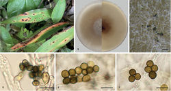
Asexual morph: Sporulated on PDA, mycelium consisting of hyaline, smooth, branched, septate hyphae 1.0–5.2 µm in diam. (n = 20). Conidiophores reduced to conidiogenous cells. Conidiogenous cells aggregated in clusters on hyphae, hyaline to pale brown, smooth, doliiform, clavate to ampulliform, 2.2–11.9 × 2.2–4.9 µm, mean (± SD): 6.4 (± 2.5) × 3.4 (± 0.6) µm (n = 50). Conidia globose, subglobose to lenticular, with a longitudinal germ slit, olive to dark brown, smooth to finely roughened and two or more conidia are produced on each conidiogenous cell, 5.1–8.9 × 4.6–7.7 µm, mean (± SD): 6.5 (± 0.8) × 5.9 (± 0.7) µm, L/W = 1.0–1.4 (n = 50). Sexual morph: Undetermined.
Culture characteristics
On PDA, colonies flat, spreading, margin circular, thick, concentrically spreading with aerial mycelium, surface light greyish-brown, reverse tawny pigment diffused in media, mycelia white to grey and pale brown, sporulation on hyphae, reaching 9 cm in 7 days at 25 °C.
Notes
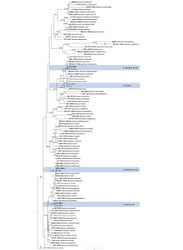
Phylogenetic analysis indicated that Apiospora lophatheri is closely related to a clade comprising A. chromolaenae, A. euphorbiae, A. italicum, A. malaysiana, A. phyllostachydis, A. thailandica and A. vietnamense (Fig. 1). We compared the new species with phylogenetically similar taxa, based on morphological differences (Table 3) and base pair differences (Table 4). A. lophatheri can be differentiated from A. chromolaenae by its wider conidiogenous cells (2.2–11.9 × 2.2–4.9 µm vs. 6.5–12 × 1–2 µm) (from Euphorbia sp.; collected in Zambia; Ellis (1965)[1]) and by 18 gene base pair differences (17/529 in ITS, 1/838 in LSU). A. lophatheri differs from A. euphorbiae by its larger olive to dark brown conidia (5.1–8.9 × 4.6–7.7 µm vs. 4–5.5 × 3–4 µm) (from Euphorbia sp.; collected in Zambia; Ellis (1965)[1]), with nucleotide differences in ITS as 3/529, in LSU as 2/318, in tub2 as 22/801. A. italicum has smaller conidia (4–6 × 3–4 µm) (from Arundo donax; collected in Italy; Pintos et al. (2019)[2]) and has 125 nucleotides differences (41/552 in ITS, 2/828 in LSU, 27/432 in tef1, 55/838 in tub2). Additionally, A. lophatheri is distinguished from A. malaysiana by having larger globose or subglobose conidia (5.1–8.9 × 4.6–7.7 µm vs. 5–6 × 3–4 µm) (from Macaranga hullettii; collected in Malaysia; Crous and Groenewald (2013)[3]), with 43 nucleotide differences (3/529 in ITS, 1/838 in LSU, 18/424 in tef1, 21/801 in tub2). A. lophatheri differs from A. phyllostachydis by its relatively shorter conidiogenous cells (2.2–11.9 × 2.2–4.9 µm vs. 20–55 × 1.5–2.5 µm) (from Phyllostachys heteroclada; collected in China; Yang et al. (2019)[4]) and by 48 nucleotides differences (7/529 in ITS, 3/838 in LSU, 12/424 in tef1, 26/795 in tub2). A. lophatheri can be differentiated from A. thailandica by having shorter conidiogenous cells (2.2–11.9 × 2.2–4.9 µm vs. 11.5–39 × 2–3.5 µm) (from bamboo; collected in Thailand; Dai et al. (2017)[5]) and by 12 nucleotides differences (9/529 in ITS, 3/828 in LSU). The conidia of A. lophatheri are significantly wider and paler-coloured than those of A. vietnamense (5.1–8.9 × 4.6–7.7 µm vs. 5–6 × 3–4 µm) (from Citrus sinensis; collected in Vietnam; Wang et al. (2018)[6]) and there are 7 nucleotides differences between the two species (2/526 in ITS, 2/803 in LSU, 3/315 in tub2). Therefore, A. lophatheri is described as a new species, based on phylogeny and morphological comparison.
| Species | Isolation source | Country | Conidiogenous cells (µm) | Conidia in surface view | Conidia in side view | References | ||
|---|---|---|---|---|---|---|---|---|
| Shape | Diam (μm) | Shape | Diam (μm) | |||||
| A. gaoyouense | Phragmites australis | China | 1–2 × 2–3 | globose to elongate ellipsoid | 5–8 | lenticular | 4–8 | Jiang et al. (2018)[7] |
| A. hispanicum | Maritime sand | Spain | – | globose to ellipsoid | 7.5–8.5 × 6–7.5 | lenticular | 6.5 | Larrondo (1992) |
| A. locuta-pollinis | Brassica campestris | China | 3–7.5 × 3–6 | globose to elongate ellipsoid | 8–15× 5–9.5 | – | – | Zhao et al. (2018)[8] |
| A. longistroma | Bamboo | Thailand | – | asexual morph: Undetermined | – | – | – | Dai et al. (2017)[5] |
| A. marii | Beach sand/ Poaceae | Spain | 5–10 × 3–4.5 | globose to elongate ellipsoid | 8–10(−13) | lenticular | (5–)6(−8) | Crous and Groenewald (2013)[3] |
| A. mediterranei | Airborn spore/ grass | Spain | – | lentiform | 9–9.5 × 7.5–9 | – | – | Larrondo (1992) |
| A. oenotherae | Oenothera biennis | China | 2.0–14.2 × 1.1–4.9 | globose, subglobose to lenticular | 6.6–13.9 × 5.5–10.1 | – | – | This study |
| A. piptatheri | Piptatherum miliaceum | Spain | 6–27 × 2–5 | globose to elongate ellips oid | 6–8 × 3–5 | lenticular | 4.5–6 | Pintos et al. (2019)[2] |
| A. pseudomarii | Aristolochia debilis | China | 8–13 × 2.5–5 | subglobose to ellipsoid | 6–9 × 4.5–6 | – | – | Chen et al. (2021)[9] |
| A. chromolaenae | Chromolaena odorata | Thailand | 6.5–12 × 1–2 | elongated, broadly fliform to ampulliform | 4–6×4.5–6.5 | – | – | Mapook et al. (2020)[10] |
| A. euphorbiae | Bambusa | Bangladesh | – | circular or nearly circular | (4–)4.7(–5.5) | lenticular | (3–)3.2(–4) | Sharma et al. (2014)[11] |
| A. italicum | Arundo donax | Italy | (3–)4–7(–9) × (1.5–)2–3(–5) | globose | 4–6×3–4 | lenticular | – | Pintos et al. (2019)[2] |
| A. lophatheri | Lophatherum gracile | China | 2.2–11.9 × 2.2–4.9 | globose, subglobose to lenticular | 5.1–8.9 × 4.6–7.7 | – | – | This study |
| A. malaysiana | Macaranga hullettii | Malaysia | 4–7 × 3–5 | globose | 5–6 | lenticular | 3–4 | Crous and Groenewald (2013)[3] |
| A. phyllostachydis | Phyllostachys heteroclada | China | 20–55 × 1.5–2.5 | globose to subglobose, oval or irregular | 5–6 × 4–6 | – | – | Yang et al. (2019)[4] |
| A. thailandicum | Bamboo | Thailand | 11.5–39 × 2–3.5 | globose to subglobose, elongated to ellipsoidal | 5–9 × 5–8 | – | – | Dai et al. (2017)[5] |
| A. vietnamense | Citrus sinensis | Vietnam | 4–7 × 3–5 | globose | 5–6 | lenticular | 3–4 | Wang et al. (2017)[12] |
| Taxa | Loci | Nucleotides difference without gaps | Rates of base pair differences |
|---|---|---|---|
| A. chromolaenae | ITS | 17/529 (40, 102, 108, 109, 110, 111, 112, 113, 114, 115, 116, 117, 118, 119, 120, 121, 122) | 3.21% |
| LSU | 1/838 (426) | 0.12% | |
| A. euphorbiae | ITS | 3/515 (26, 88, 89) | 0.58% |
| LSU | 2/318 (146, 306) | 0.63% | |
| tub2 | 22/801 (95, 96, 123, 151, 154, 163, 166, 182, 185, 193, 216, 237, 312, 347, 372, 429, 453, 454, 474, 559, 569, 574) | 2.75% | |
| A. italicum | ITS | 41/552 (40, 82, 93, 94, 95, 96, 97, 98, 99, 100, 101, 102, 103, 104, 105, 106, 107, 108, 109, 110, 111, 112, 113, 114, 115, 116, 117, 118, 119, 120, 121, 122, 132, 165, 177, 180, 205, 207, 213, 487, 529) | 7.43% |
| LSU | 2/828 (406, 416) | 0.24% | |
| tef1 | 27/432 (16, 18, 19, 20, 21, 22, 23, 24, 25, 27, 35, 46, 53, 60, 75, 80, 90, 102, 119, 123, 125, 172, 210, 211, 240, 248, 272) | 6.25% | |
| tub2 | 55/838 (5, 29, 44, 45, 46, 92, 99, 119, 121, 122, 126, 155, 157, 171, 185, 188, 193, 194, 196, 198, 202, 297, 219, 229, 240, 265, 315, 338, 358, 363, 367, 368, 382, 384, 386, 390, 403, 407, 412, 430, 434, 454, 463, 465, 467, 480, 491, 499, 502, 556, 564, 580, 642, 756, 757) | 6.56% | |
| A. malaysiana | ITS | 3/529 (40, 102, 103) | 0.57% |
| LSU | 1/838 (426) | 0.12% | |
| tef1 | 18/424 (15, 16, 19, 27, 29, 38, 52, 56, 82, 83, 91, 93, 95, 111, 115, 202, 203, 264) | 4.25% | |
| tub2 | 21/801 (95, 96, 123, 151, 154, 163, 166, 182, 185, 193, 216, 237, 312, 347, 372, 429, 453, 474, 559, 569, 574) | 2.62% | |
| A. phyllostachydis | ITS | 7/529 (40, 44, 85, 102, 106, 433, 500) | 1.32% |
| LSU | 3/838 (7,8,9) | 0.36% | |
| tef1 | 12/424 (16, 19, 26, 27, 51, 52, 53, 111, 197, 202, 203, 264) | 2.83% | |
| tub2 | 26/795 (35, 52, 55, 84, 89, 112, 116, 147, 151, 175, 178, 186, 209, 211, 231, 329, 352, 354, 360, 462, 469, 489, 570, 572, 575, 608) | 3.27% | |
| A. thailandicum | ITS | 9/529 (40, 82, 102, 107, 122, 175, 177, 183, 501) | 1.70% |
| LSU | 3/828 (5, 416, 434) | 0.36% | |
| A. vietnamense | ITS | 2/526 (37, 99) | 0.38% |
| LSU | 2/803 (237, 391) | 0.25% | |
| tub2 | 3/315 (72, 82, 87) | 0.95% |
Original Description
- Li, S; Peng, C; Yuan, R; Tian, C; 2023: Morphological and phylogenetic analyses reveal three new species of Apiospora in China MycoKeys, 99: 297-317. doi
Images
|
Other References
- ↑ 1.0 1.1 Ellis M (1965) Dematiaceous Hyphomycetes. VI.Mycological Papers103: 1–46.
- ↑ 2.0 2.1 2.2 Pintos Á, Alvarado P, Planas J, Jarling R (2019) Six new species of Arthrinium from Europe and notes about A. caricicola and other species found in Carex spp. hosts.MycoKeys49: 15–48. https://doi.org/10.3897/mycokeys.49.32115
- ↑ 3.0 3.1 3.2 Crous P, Groenewald J (2013) A phylogenetic re-evaluation of Arthrinium.IMA Fungus4(1): 133–154. https://doi.org/10.5598/imafungus.2013.04.01.13
- ↑ 4.0 4.1 Yang C, Xu X, Dong W, Wanasinghe D, Liu Y, Hyde K (2019) Introducing Arthrinium phyllostachium sp. nov. (Apiosporaceae, Xylariales) on Phyllostachys heteroclada from Sichuan province, China.Phytotaxa406(2): 91–110. https://doi.org/10.11646/phytotaxa.406.2.2
- ↑ 5.0 5.1 5.2 Dai D, Phookamsak R, Wijayawardene N, Li W, Bhat D, Xu J, Taylor J, Hyde K, Chukeatirote E (2017) Bambusicolous fungi.Fungal Diversity82(1): 1–105. https://doi.org/10.1007/s13225-016-0367-8
- ↑ Wang M, Tan X, Liu F, Cai L (2018) Eight new Arthrinium species from China.MycoKeys1: 1–24. https://doi.org/10.3897/mycokeys.39.27014
- ↑ Jiang N, Li J, Tian C (2018) Arthrinium species associated with bamboo and reed plants in China.Fungal Systematics and Evolution13: 217–229. https://doi.org/10.3114/fuse.2018.02.01
- ↑ Zhao Y, Zhang Z, Cai L, Peng W, Liu F (2018) Four new filamentous fungal species from newly-collected and hive-stored bee pollen.Mycosphere: Journal of Fungal Biology9(6): 1089–1116. https://doi.org/10.5943/mycosphere/9/6/3
- ↑ Chen T, Zhang Y, Ming X, Zhang Q, Long H, Hyde K, Li Y, Wang Y (2021) Morphological and phylogenetic resolution of Arthrinium from medicinal plants in Yunnan, including A. cordylines and A. pseudomarii spp. nov.Mycotaxon136(1): 183–199. https://doi.org/10.5248/136.183
- ↑ Mapook A, Hyde K, McKenzie E, Bhat D, Jeewon R, Stadler M, Samarakoon M, Malaithong M, Tanunchai B, Buscot F, Wubet T, Purahong W (2020) Taxonomic and phylogenetic contributions to fungi associated with the invasive weed Chromolaena odorata (Siam weed).Fungal Diversity101(1): 1–175. https://doi.org/10.1007/s13225-020-00444-8
- ↑ Sharma R, Kulkarni G, Sonawane M, Shouche Y (2014) A new endophytic species of Arthrinium (Apiosporaceae) from Jatropha podagrica.Mycoscience55(2): 118–123. https://doi.org/10.1016/j.myc.2013.06.004
- ↑ Wang M, Liu F, Crous P, Cai L (2017) Phylogenetic reassessment of Nigrospora: Ubiquitous endophytes, plant and human pathogens.Persoonia39(1): 118–142. https://doi.org/10.3767/persoonia.2017.39.06

