Apiospora arundinis
| Notice: | This page is derived from the original publication listed below, whose author(s) should always be credited. Further contributors may edit and improve the content of this page and, consequently, need to be credited as well (see page history). Any assessment of factual correctness requires a careful review of the original article as well as of subsequent contributions.
If you are uncertain whether your planned contribution is correct or not, we suggest that you use the associated discussion page instead of editing the page directly. This page should be cited as follows (rationale):
Citation formats to copy and paste
BibTeX: @article{Li2023MycoKeys99, RIS/ Endnote: TY - JOUR Wikipedia/ Citizendium: <ref name="Li2023MycoKeys99">{{Citation See also the citation download page at the journal. |
Ordo: Xylariales
Familia: Apiosporaceae
Genus: Apiospora
Name
Apiospora arundinis (Corda) Pintos & P. Alvarado, Fungal Systematics and Evolution 7: 205 (2021) – Wikispecies link – Pensoft Profile
Description
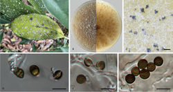
Asexual morph: Mycelium consisting of smooth, hyaline, branched, septate, 1.1–5.9 µm diam. hyphae (n = 20). Conidiophores reduced to conidiogenous cells. Conidiogenous cells subglobose to ampulliform, erect, blastic, aggregated in clusters on hyphae, smooth, branched, 3.4–9.4 × 1.5–6.4 µm, mean (± SD): 6.8 (± 1.6) × 3.9 (± 1.3) µm (n = 50). Conidia globose, subglobose to lenticular, with a longitudinal germ slit, occasionally elongated to ellipsoidal, brown to dark brown, smooth to finely roughened, 6.4–10.4 × 5.2–8.3 µm, mean (± SD): 7.7 (± 0.6) × 6.8 (± 0.7) µm, L/W = 1.0–1.5 (n = 50). Sexual morph: Undetermined.
Culture characteristics
On PDA, colonies thick and dense, margin undulate and irregular, pale yellow pigment diffused into medium, surface with patches of iron-grey aerial mycelia, reverse yellowish-brown, mycelia white to grey, sporulation on hyphae, reaching 9 cm in 7 days at 25 °C.
Specimens examined
China, Yunnan Province: Xishuangbanna Botanical Garden, on diseased leaves of Brunfelsia brasiliensis, 6 June 2022, S.J. Li, BJFC-S1918; living cultures CFCC 58977, LS 107).
Notes
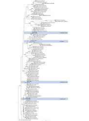
In this study, two isolates clustered together with the culture of A. arundinis with high-support values (ML/BI = 100/0.99)in the multi-locus phylogenetic tree (Fig. 1). Thus, these isolates were identified as A. arundinis and Brunfelsia brasiliensis as a new host record for this species. Apiospora arundinis was introduced from Phyllostachys praecox, Castanea mollissima and Saccharum officinarum in China (Chen et al. 2014[1]; Jiang et al. 2021[2]; Liao et al. 2022[3]). Comparing with the description from Chen et al. (2014)[1] (5–7 × 2–4 µm), Jiang et al. (2021)[2] (3–4 µm) and Liao et al. (2022)[3] (4.5–7.4 × 3.3–4.4 µm), the conidia in this study show larger sizes (6.4–10.4 × 5.2–8.3 µm). These differences may result from different host and habitat.
Taxon Treatment
- Li, S; Peng, C; Yuan, R; Tian, C; 2023: Morphological and phylogenetic analyses reveal three new species of Apiospora in China MycoKeys, 99: 297-317. doi
Images
|
Other References
- ↑ 1.0 1.1 Chen K, Wu X, Huang M, Han Y (2014) First report of brown culm streak of Phyllostachys praecox caused by Arthrinium arundinis in Nanjing, China. Plant Disease 98(9): е1274. https://doi.org/10.1094/PDIS-02-14-0165-PDN
- ↑ 2.0 2.1 Jiang N, Fan X, Tian C (2021) Identification and characterization of leaf-inhabiting fungi from Castanea plantations in China.Journal of Fungi7(1): 1–64. https://doi.org/10.3390/jof7010064
- ↑ 3.0 3.1 Liao J, Jiang W, Wu X, He J, Li H, Wang T, Cheng L, Chen W, Mo L (2022) First report of Apiospora mold on Sugarcane in China caused by Apiospora arundinis (Arthrinium arundinis). Plant Disease 106(3): е1058. [Epub2022Feb16] https://doi.org/10.1094/PDIS-02-21-0386-PDN

