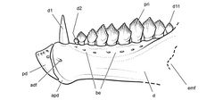
Pegomastax africanus
| Notice: | This page is derived from the original publication listed below, whose author(s) should always be credited. Further contributors may edit and improve the content of this page and, consequently, need to be credited as well (see page history). Any assessment of factual correctness requires a careful review of the original article as well as of subsequent contributions.
If you are uncertain whether your planned contribution is correct or not, we suggest that you use the associated discussion page instead of editing the page directly. This page should be cited as follows (rationale):
Citation formats to copy and paste
BibTeX: @article{Sereno2012ZooKeys226, RIS/ Endnote: TY - JOUR Wikipedia/ Citizendium: <ref name="Sereno2012ZooKeys226">{{Citation See also the citation download page at the journal. |
Ordo: Ornithischia
Familia: Heterodontosauridae
Genus: Pegomastax
Name
Pegomastax africanus Sereno, 2012 sp. n. – Wikispecies link – ZooBank link – Pensoft Profile
Holotype
SAM-PK-K10488, fragmentary skull preserving right and left dentaries and the predentary.
Type locality
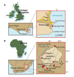
Voyizane (= Voisana), Transkei (Herschel) District, Cape Province, South Africa; S30°34', E27°25' (Crompton and Charig 1962[1]; Kitching and Raath 1984[2]) (Fig. 1B).
Horizon.Upper section of the Elliot Formation (= Red Beds); Lower Jurassic, Hettangian to Sinemurian, ca. 200-190 Ma (Crompton and Charig 1962[1]; Santa Luca et al. 1976[3]; Knoll 2005[4]; Gradstein and Ogg 2009[5]).
Derivation of name
From “Latin africanus, meaning “pertaining to Africa”.
Diagnosis
Heterodontosaurid ornithischian characterized by the following four autapomorphies: (1) proportionately deep predentary with a dorsal margin about 70% the anteroventral margin; (2) predentary dorsal margin angled anteroventrally at approximately 45°; (3) postcaniniform dentary crowns with an mesially-bowed primary ridge that angles from the apical denticle toward the mesial side of the crown base; (4) slightly concave denticulate crown margins to either side of a prominent apical denticle.
Description
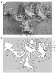
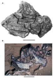
The holotypic represents a small heterodontosaurid that is preserved on a small block of sandstone matrix (Figs 5B, 82) collected at Voyizane (Fig. 1B) near sites that yielded the best-preserved remains of Heterodontosaurus tucki (SAM-PK-K337, -K1332). Only portions of the skull are preserved, which includes the right postorbital, both dentaries, and the predentary. Further preparation or computed-tomographic imaging may allow identification of other elements. The anterior end of the lower jaws is preserved close to their natural articulation. The left dentary has slid slightly ventral to the right dentary, exposing a portion of its symphyseal articular surface (Figs 82–84). Bones that should be present medial to the dentaries, such as the coronoid and splenial, have moved from their natural articulation if they are preserved.
Predentary
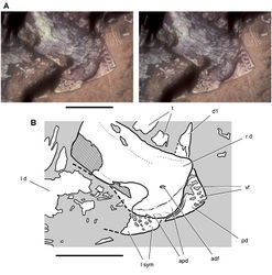
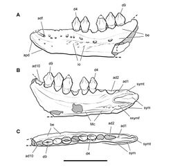
The predentary is a wedge-shaped bone that is very short anteroposteriorly (Figs 84, 87). Its dorsal margin is only about 70% of the dorsoventral depth of the bone. In Heterodontosaurus, in contrast, the dorsal margin of the predentary slightly exceeds its dorsoventral depth (Figs 59-61). Matrix currently fills several vascular foramina and associated grooves below the sharp dorsal margin of the predentary, which would have supported a keratinous bill (Fig. 84). The row of foramina parallels the sharp dorsal edge of the bone, which shows no evidence of breakage despite its strong anteroventral inclination at about 45°. The inclination of the dorsal margin of the predentary appears natural, as the bone is preserved in articulation with its dorsal edge butted against the right dentary. The posterior margin of the predentary is sinuous, tapering ventrally to a point among the midline (Fig. 84). As in Heterodontosaurus, there is no development of a discrete lateral predentary process, and the posterior margin is positioned against the upper part of a saddle-shaped articular surface along the anterior end of the dentary.
Dentary
The robustly proportioned right and left dentaries are exposed in lateral and medial views, respectively (Figs 83, 84, 87). The dentary tooth row has a length of approximately 27 mm, as measured from the anterior end of the left dentary to the posterior edge of its posteriormost dentary crown (Fig. 82). The depth of the dentary ramus at mid-length is approximately 9 mm, or about one-third of the total length of the tooth row. In Heterodontosaurus, in contrast, the dentary tooth row has a length of 42 mm and a depth at mid-length of 10 mm (SAM-PK-K1332), for relative depth less than one-quarter the length of the toothed portion of the bone. The dentary in Manidens (Fig. 81B) appears to be slightly more robust than that in Pegomastax by this proportion.
The lateral aspect of the right dentary preserves a deep buccal emargination including several matrix-filled foramina. A small anterior dentary foramen is present near the predentary and is not associated with an impressed vessel tract as in Echinodon and Fruitadens (Figs 9A, 16). The blunt anterior end of the dentary has a smooth, saddle-shaped articular surface for the predentary, which is narrower dorsally and broadest ventral to the midline. Although fitted to the sinuous posterior margin of the predentary, the articular surface is significantly deeper than the predentary, similar to the condition in Heterodontosaurus (Fig. 39). The dorsal end of the saddle-shaped articular surface extends under the predentary near the prominent anterodorsal corner of the dentary. The ventral end of the trough-shaped articulation is situated below the ventral margin of the dentary ramus. The anterior end of the dentary is expanded dorsoventrally relative to the main body of the dentary ramus (Fig. 87), as in Abrictosaurus and Heterodontosaurus.
The flat ventral portion of the dentary symphysis is exposed in medial view of the left dentary (Fig. 84). Meckel’s canal is exposed as a narrow trough located just above the ventral margin of the left dentary ramus as in other heterodontosaurids (Fig. 82). A robust tongue-shaped coronoid process rises at about 45° from the posterior end of the tooth row. Replacement foramina do not appear to be present near the alveolar margin of the left dentary, which is well exposed in medial view.
Postorbital
The right postorbital is preserved in medial view (Fig. 82). The most notable feature is the deep proportions of the posterior ramus, which resembles the condition in Manidens (Fig. 81) in contrast to the more slender ramus in Heterodontosaurus (Fig. 59). The arched ventral ramus forms the posterior portion of the orbital margin.
Dentary teeth
There are probably 11 dentary teeth. Most of the right tooth row is preserved in lateral view from the caniniform tooth (d1) to d8 (Fig. 82). The left dentary preserves the coronoid process and five relatively large crowns at the posterior end of the tooth row (d7-11). The longer length of the dentigerous portion of the left dentary, from its anterior end to the posteriormost crown, suggests that the posterior portion of the right dentary and several posterior teeth have been sheared away. This is confirmed on the breakage surface of the right dentary, where the roots of three posterior teeth are seen in the cross-section (Figs 82, 83). The right dentary, thus, suggests there are at least 11 teeth in the dentary, and that the left dentary crowns represent d7-11 (Fig. 86). The anterior portion of the left dentary tooth row may be preserved but obscured by the right dentary.
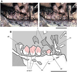
The caniniform tooth (d1) has straight mesial and distal carinae, the former with serrations (Fig. 85). The base of the crown is crushed and the upper one-half slightly twisted. The distal carina was probably straight like the mesial carina. Unlike the mesial carina, the distal carina does not appear to have had serrations. The lack of any distal recurvature in the crown of the dentary caniniform tooth contrasts with the condition in most heterodontosaurids including Lycorhinus, Abrictosaurus and Heterodontosaurus.
There is a significant diastema in the right dentary tooth row between the caniniform tooth and d2, the relatively small first postcaniniform crown (Fig. 85, 87). Right d2-4 show an increase in crown size, and crown size seems to culminate in d6 (Fig. 85). The crowns of d2 and d3 are diamond shaped with a basal constriction under the crown. Like all dentary crowns, they are asymmetrical, with the distal apical margin slightly longer and extending farther down the crown than the mesial apical margin. A few denticles are preserved on their margins but these are not well preserved. The crown of d5 is diamond-shaped with six or seven denticles preserved distal to the apical denticle. Like more distal crowns, most of its lingual face is worn away by a planar wear facet, which has yet to obliterate the distal denticles. Small areas of the crown surface of d3 and d4 have a glassy appearance, suggesting that a thin layer of enamel may have been retained on the labial side of the dentary crowns.
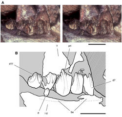
Right d6 and d7 have a dorsal crown profile like all more distal crowns. Concave apical margins are present on either side of the apical denticle, the mesial apical margin of which is shorter and more apically positioned than the distal apical margin (Fig. 86). Adjacent worn crowns create a scalloped leading edge, as preserved in the middle of the right tooth row (Fig. 85). The medial crowns surface is most completely preserved in distal teeth (d7-11) on the left side (Fig. 86). All but the distalmost corner of the crown of d9 is exposed, which has a height 150% of its maximum width. The crown expands above its root, although the crown-root junction in this tooth is not exposed. The margins of the basal portion of the crown are not raised, and the first denticle on the apical margin on either side is not enlarged or divergent as in Manidens. Pegomastax has a well-developed primary ridge that originates just above the crown base and gains prominence as it arches from the mesial to the center of the crown before joining the apical denticle distal to the central crown axis (Fig. 86). The terminal end of the ridge is wide enough that it incorporates small accessory denticles, one to each side of the apical denticle, an unusual feature that may only be expressed in the largest crowns. There are about eight denticles to each side of the apical triumvirate. The denticles appear to be slightly larger on the distal apical margin, which is slightly longer and more steeply inclined from the horizontal than the mesial apical margin. The distalmost dentary tooth (d11) has lower proportions, as is typical for the last crown of the tooth row in heterodontosaurids. Its crown is only slightly taller than it is wide (Fig. 86).
The roots are only partially exposed in cross-section of the right lower jaw. The most complete of these, which probably belongs to d10, has a relatively large diameter and central lumen. The roots in Pegomastax thus may have been swollen as in many other heterodontosaurids.
The crowns of right d5-8 and left d6-9 have an imbricate arrangement relative to one another (Figs 85-87). This feature is common in ornithischian dentitions and positions the mesial edge of each crown lingual to the distal edge of the next most mesial crown. The distal two-thirds of the dentary tooth row in Pegomastax, thus, exhibits this minor overlap between adjacent crowns edges. There does not appear to be a mesial fossa at the base of the crown to accommodate the edge of an adjacent crown as in Manidens (Pol et al. 2011[6]). This fossa in Manidens, however, is only well exposed in mesial view, a view not yet available in Pegomastax.
All postcaniniform teeth in the right dentary are worn to varying degrees except perhaps the small crowns of d2-4 (Fig. 85). Sustained wear has obliterated all denticles on d6 and d7 in the right dentary. On the left side, d10 has reached this stage of wear and has no denticles; right d7 and d8 show less wear, and d9 and d11 show little or no wear. Their broad, nearly planar wear surfaces join to form a nearly continuous shearing surface, which is set at a very low-angle to the vertical crown axis.
Differential wear along the tooth row strongly suggests cyclic tooth replacement, despite the general alignment of wear surfaces and the absence of replacement foramina. Direct evidence of replacement is present in the left dentary tooth row. A small replacement crown is emerging at the base of the heavily worn crown of d10 (Fig. 86).
Jaw reconstruction
The reconstruction of the dentary of Pegomastax is based on the holotype and only known specimen (Fig. 87). The labial surfaces of available dentary crowns are worn or broken, obscuring details of ornamentation. In the skull reconstruction, the ornamentation of labial crown surfaces was tentatively based on the exposed lingual crown surfaces in the left dentary (Fig. 86). Computed tomographic imaging might reveal evidence of the form of the lateral crown surface in a relatively unworn crown such as left d9 (Fig. 86).
The most unusual feature of the preserved portion of the skull is the shape of the predentary. Despite many similarities to Heterodontosaurus in the differentiated dentition and the saddle-shaped form of the predentary-dentary articular surface, the unusually deep, parrot-like shape of the predentary suggests that its function in cropping vegetation may have become specialized in various heterodontosaurid species, similar to the way bill shape varies among birds. Pegomastax, furthermore, provides additional evidence in favor of a mobile predentary-dentary joint, given the disparity in size between the predentary and the longer articular surface on the dentaries.
Original Description
- Sereno, P; 2012: Taxonomy, morphology, masticatory function and phylogeny of heterodontosaurid dinosaurs ZooKeys, 226: 1-225. doi
Images
|
Other References
- ↑ 1.0 1.1 Crompton A, Charig A (1962) A new ornithischian from the Upper Triassic of South Africa. Nature 196: 1074-1077. doi: 10.1038/1961074a0
- ↑ Kitching J, Raath M (1984) Fossils from the Elliot and Clarens Formations (Karoo Sequence) of the northeastern Cape, Orange Free State and Lesotho, and a suggested biozonation based on tetrapods. Palaeontologia africana 25: 111-125.
- ↑ Santa Luca A, Crompton A, Charig A (1976) A complete skeleton of the Late Triassic ornithischian Heterodontosaurus tucki. Nature 264: 324-328.
- ↑ Knoll F (2005) The tetrapod fauna of the Upper Elliot and Clarens formations in the main Karoo Basin (South Africa and Lesotho). Bulletin de la Société géologique de France 176: 81-91. doi: 10.2113/176.1.81
- ↑ Gradstein F, Ogg J (2009) The geologic time scale. In: Hedges S Kumar S (Eds). , The Timetree of Life. Oxford University Press, Oxford: 26-34.
- ↑ 6.0 6.1 Pol D, Rauhut O, Becerra M (2011) A Middle Jurassic heterodontosaurid dinosaur from Patagonia and the evolution of heterodontosaurids. Naturwissenschaften 98: 369-379.
- ↑ Butler R, Galton P, Porro L, Chiappe L, Henderson DM et a (2010) Lower limits of ornithischian dinosaur body size inferred from a new Upper Jurassic heterodontosaurid from North America. Proceedings of the Royal Society B: Biological Sciences 277: 375-381. doi: 10.1098/rspb.2009.1494
- ↑ Zheng X, You H, Xu X, Dong Z (2009) Early Cretaceous heterodontosaurid dinosaur with integumentary structures. Nature 458: 333–336. doi: 10.1038/nature07856
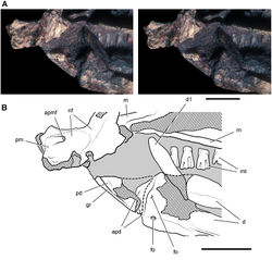
![Figure 9. More recent heterodontosaurid discoveries from northern locales. A Jaws of Fruitadens haagarorum from the Upper Jurassic Morrison Formation in Colorado, USA (based on LACM 115747, 128258; reversed from Butler et al. 2010[7]) B Left dentary in lateral view of an undescribed heterodontosaurid from the Lower Jurassic Kayenta Formation of Arizona (from Sereno et al. unpublished) C Partial skull of Tianyulong confuciusi from the Yixian Formation of Liaoning Province, PRC (STMN 26-3; reversed from Zheng et al. 2009[8]). Abbreviations: a angular ad 9, 10 alveolus for dentary tooth 9, 10 adf anterior dentary foramen antfo antorbital fossa apd articular surface for the predentary d dentary d1, 2, 8 dentary tooth 1, 2, 8 emf external mandibular fenestra en external nares j jugal l lacrimal m maxilla n nasal pd predentary pf prefrontal pm premaxilla po postorbital q quadrate qj quadratojugal sa surangular. Scale bar equals 1 cm in A and C and 5 mm in B.](https://species-id.net/o/thumb.php?f=ZooKeys-226-001-g009.jpg&width=201)
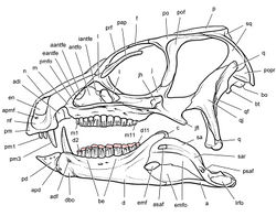

![Figure 81. Partial skull of Manidens condorensis from the Middle Jurassic Cañadón Asfalto Formation of Argentina. Skull reconstructions in lateral view A Reversed from Pol et al. (2011)[6] B This study. Dashed lines indicate estimated edges. Abbreviations: a angular antfo antorbital fossa asaf anterior surangular foramen be buccal emargination bo basioccipital bt basal tubera d dentary d1, 2, 11 dentary tooth 1, 2, 11 emfo external mandibular fossa f frontal gl glenoid gr groove j jugal jfl jugal flange jh jugal horn m maxilla m1, 11 maxillary tooth 1, 11 n nasal pd predentary pm premaxilla po postorbital pof postorbital fossa popr paroccipital process q quadrate qj quadratojugal ri ridge sa surangular sq squamosal.](https://species-id.net/o/thumb.php?f=ZooKeys-226-001-g081.jpg&width=235)
