Difference between revisions of "Ortheziola viti"
m (Imported from ZooKeys) |
m (1 revision) |
(No difference)
| |
Latest revision as of 13:05, 30 April 2014
| Notice: | This page is derived from the original publication listed below, whose author(s) should always be credited. Further contributors may edit and improve the content of this page and, consequently, need to be credited as well (see page history). Any assessment of factual correctness requires a careful review of the original article as well as of subsequent contributions.
If you are uncertain whether your planned contribution is correct or not, we suggest that you use the associated discussion page instead of editing the page directly. This page should be cited as follows (rationale):
Citation formats to copy and paste
BibTeX: @article{Kaydan2014ZooKeys406, RIS/ Endnote: TY - JOUR Wikipedia/ Citizendium: <ref name="Kaydan2014ZooKeys406">{{Citation See also the citation download page at the journal. |
Ordo: Hemiptera
Familia: Coccoidea
Genus: Ortheziola
Name
Ortheziola viti Szita & Konczné Benedicty sp. n. – Wikispecies link – ZooBank link – Pensoft Profile
Material examined
Holotype. Adult female. Greece, Thessaly, North-Tsakgarada, 550 m a.s.l., in hollow base of Platanus sp., 09 Apr 2004, Leg. S. Vit [MHNG: GR-2004 No.3; PPI code: 8915].
Paratypes. 2 adult females, 1 specimen on same slide as holotype, 1 specimen on separate slide, same data as holotype [MHNG code: GR-2004 No.3; PPI code: 8915]; 4 females on 3 slides: Turkey, North Elma-Dagi, Ankara-slopes, 1200 m a.s.l., Crataegus sp. litter, 31 Oct 1995 Leg. S. Vit [MHNG code: ANK. No.3; PPI code: 9463].
Description
Unmounted adult female. Not seen.
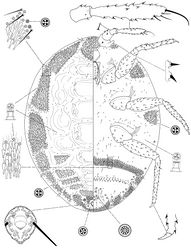
Slide mounted adult female. Body 1.320–1.476 mm; 1.030–1.192 mm wide. Length of antennal segments: 1st 60–75 µm; 2nd 36–51 µm; 3rd 208–228 µm; 3rd segment parallel sided or weakly clubbed; apical seta 91–120 µm long, subapical seta 32–40 µm long; fleshy sensory seta near apical seta 11–14 µm long; microseta present near apex of antenna; unusual hair-like seta present near subapical seta; all segments of antennae covered with moderate number of spine-like, straight, apically acute setae, longest seta 10 µm long; first antennal segment with one seta on each side of segment.
Venter. Labium 117–136 µm long. Stylet loop about as long as labium. Leg segment lengths: front coxa 90–102 µm, middle 97–107 µm, hind 120–140 µm; front trochanter-femur 235–244 µm, middle 240–276 µm, hind 281–302 µm; front tibia-tarsus 250–269 µm, middle 269–282 µm, hind 326–355 µm; front claw 40–47 µm, middle 40–44 µm, hind 44–48 µm long; claw digitules spine-like, 5–6.5 µm long; legs with rows of robust setae; longest seta on trochanter-femur 10–12 µm; with one flagellate sensory seta on tibia, 21 µm long; each trochanter with 4 sensory sensilla on each surface. Wax plates absent from marginal areas of head and thorax except for small spine cluster next to antenna (plate 12) and normal plate between antennae (plate 11), and with marginal wax band surrounding each thoracic spiracle (plates 15 and 16); without triangular-shaped wax plates in front of coxae (plates 13, 17 and 18); plate 19 absent; without cluster of spines between hind legs and ovisac band; anterior edge of ovisac band almost completely straight; with one band of spines within ovisac band. Thoracic spiracles each with scattered quadrilocular pores loosely associated with spiracle opening, each group containing 20–28 pores, each pore 4 µm in diameter (several of these pores present on dorsum); diameter of opening of anterior thoracic spiracle 18–22 µm. Setae few, scattered in medial areas of thorax, with several setae near anterior edge of ovisac band (some of them capitate), several associated with anterior and posterior multilocular pore rows, several more associated with posterior multilocular pores surrounding vulva. Multilocular pores each 7 µm in diameter, with 8–12 loculi around perimeter and one loculus in central hub; with quadrilocular pores predominant near anterior edge of spine band, partial row of multilocular pores near anterolateral edge of spine band, also scattered around vulva and near ovisac band, almost forming a row on the apical abdominal segment. Abdominal spiracles present with 2 pairs on each side of body anterior of ovisac band and one pair inside ovisac band, near anterolateral angle; each abdominal spiracle with sclerotized vestibule.
Dorsum. Wax plates covering two-thirds of marginal area; mediolateral thoracic plates absent (plates 3, 5 and 6); medial area of thorax and abdomen without spines and pores. Spines at margin of wax plate 4 each 17–18 µm long, in middle of wax plate each 15–18 µm long; spines truncate and expanded at apex. Flagellate setae present in marginal clusters near posterior edges of marginal wax plates (plates 2 and 4), with 2–4 setae lateral of each thoracic spiracle, each 18–20 µm long; also present in very small numbers on other wax plates and in medial bare area. Multilocular pores, each 4 µm in diameter, with 4 loculi, present in marginal areas of abdomen; also present in cluster near anal ring, the pores in this cluster sometimes each with 5 loculi. Sclerotized plate on abdomen 45–60 µm long, 190–240 µm wide; several setae situated at posterior edge of plate, many with capitate apices. Anal ring with incomplete triple row of circular pores, each pore 2–3 µm in diameter; longest anal ring seta 43–49 µm long (about equal to length of anal ring); anal ring 47–54 µm wide. Thumb-like pores each 5–6 µm long. Modified pores each 5–7 µm long. Abdominal spiracle in centre of multilocular pore cluster situated laterad of anal ring.
Host plant
Unknown.
Distribution
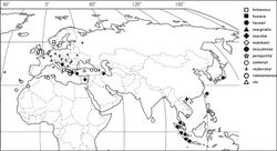
Greece, Turkey (Fig. 4).
Etymology
The new species is named after S. Vit, the collector of the type series.
Comments
Ortheziola viti is characterized by having dorsal wax plate 3 divided medially; ventral plate 19 absent from near the body margin, and dorsal plates 3, 5 and 6 absent. This species is very close to Ortheziola marginalis but differs by having (character in brackets belongs to Ortheziola marginalis): i.) short spines on antenna, the longest 10 µm (shortest 19 µm); ii.) plates on abdominal segments III-VII all divided (plates on abdominal segments III-VII not divided); and iii.) ventral plates 11 and 12 present (absent).
Original Description
- Kaydan, M; Konczné Benedicty, Z; Szita, É; 2014: New species of the genus Ortheziola Šulc (Hemiptera, Coccoidea, Ortheziidae) ZooKeys, 406: 65-80. doi
Images
|

