Ptilobactrum
| Notice: | This page is derived from the original publication listed below, whose author(s) should always be credited. Further contributors may edit and improve the content of this page and, consequently, need to be credited as well (see page history). Any assessment of factual correctness requires a careful review of the original article as well as of subsequent contributions.
If you are uncertain whether your planned contribution is correct or not, we suggest that you use the associated discussion page instead of editing the page directly. This page should be cited as follows (rationale):
Citation formats to copy and paste
BibTeX: @article{Reemer2013ZooKeys288, RIS/ Endnote: TY - JOUR Wikipedia/ Citizendium: <ref name="Reemer2013ZooKeys288">{{Citation See also the citation download page at the journal. |
Ordo: Diptera
Familia: Syrphidae
Name
Ptilobactrum Bezzi – Wikispecies link – Pensoft Profile
- Ptilobactrum Bezzi, 1915: 136. Type species: Ptilobactrum neavei Bezzi, 1915: 137, by original designation.
Description
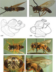
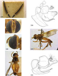
Body length: 13 mm. Broadly built flies with very wide head, long antennae and orange markings on abdomen. Head wider than thorax. Face much wider than eye; dorsally with oblique groove from lunule to eye margin; convex in profile. Lateral oral margins weakly produced. Vertex not swollen, more or less flat, but much wider than eye. Occiput narrow ventrally, moderately widened dorsally. Eye bare. Eye margins in male not converging at level of frons, with mutual distance about seven times width of antennal fossa. Antennal fossa somewhat higher than wide. Antenna longer than height of head. Basoflagellomere five times as long as scape; with long pilosity. Postpronotum pilose. Mesoscutum with transverse suture incomplete. Scutellum without calcars. Anepisternum with deep sulcus; entirely pilose. Anepimeron entirely pilose. Katepimeron convex; smooth; bare. Wing: vein R4+5 with posterior appendix; vein M1 straight, somewhat recurrent; postero-apical corner of cell r4+5 angular, with small appendix; crossvein r-m located around basal 1/3 of cell dm. Abdomen oval, widest at posterior margin of tergite 2. Tergites 3 and 4 fused. Sternite 1 bare. Sternite 4 in male visible from below. Male genitalia: phallus bent dorsad, except extreme apex bent ventrad; phallus furcate near apex; epandrium without ventrolateral ridge; surstylus broad, unfurcate, with short posterior lobe. Female unknown.
Diagnosis
Vein R4+5 with posterior appendix. Basoflagellomere with long pile. Abdomen oval. Tergites 3 and 4 fused.
Discussion
Bezzi (1915)[1] distinguished Ptilobactrum from Microdon species by the “breadth of the head, the face being furrowed, and by the unusual shape of the antennae.” Indeed, the grooves on the face, running fom the lunula obliquely downward to the eye margins, are quite unusual among Microdontinae. They are reminescent of the ptilinal sutures of Diptera Schizophora. Similar grooves are found in certain species of Furcantenna,Schizoceratomyia, Paramixogaster and Thompsodon, but usually less distinct. The antennae are unusual in their long pilosity, a character shared with Ceratrichomyia, Furcantenna, Kryptopyga and Schizoceratomyia.
See Ceratrichomyia for a discussion on synonymy of that genus with Ptilobactrum, as proposed by Cheng and Thompson (2008)[2].
Diversity and distribution
Described species: 1. Afrotropical, only known from Kenya.
Taxon Treatment
- Reemer, M; Ståhls, G; 2013: Generic revision and species classification of the Microdontinae (Diptera, Syrphidae) ZooKeys, 288: 1-213. doi
Other References
- ↑ Bezzi M (1915) The Syrphidae of the Ethiopian region. British Museum (Natural History), London, 146 pp.
- ↑ Cheng X, Thompson F (2008) A generic conspectus of the Microdontinae (Diptera: Syrphidae) with the description of two new genera from Africa and China. Zootaxa 1879: 21-48.
Images
|

