This page was moved here from http://phytopathology.net/Portal/Heterosporium_gracile
It has the following original authors (copy of the complete editing history):
(cur | prev) 18:41, 10 September 2009 Georgy Pestsov (Talk | contribs | block) . . (7,044 bytes) (0) . . (rollback 3 edits | undo)
(cur | prev) 12:36, 10 September 2009 Georgy Pestsov (Talk | contribs | block) . . (7,044 bytes) (+4) . . (undo)
(cur | prev) 15:33, 9 September 2009 Georgy Pestsov (Talk | contribs | block) . . (7,040 bytes) (-2) . . (undo)
(cur | prev) 15:29, 8 September 2009 Gregor Hagedorn (Talk | contribs | block) . . (7,042 bytes) (+65) . . (undo)
(cur | prev) 18:24, 7 September 2009 Georgy Pestsov (Talk | contribs | block) . . (6,977 bytes) (-215) . . (undo)
(cur | prev) 18:19, 7 September 2009 Georgy Pestsov (Talk | contribs | block) . . (7,192 bytes) (+200) . . (undo)
(cur | prev) 18:16, 7 September 2009 Georgy Pestsov (Talk | contribs | block) . . (6,992 bytes) (+124) . . (undo)
(cur | prev) 18:13, 7 September 2009 Georgy Pestsov (Talk | contribs | block) . . (6,868 bytes) (+123) . . (undo)
(cur | prev) 18:10, 7 September 2009 Georgy Pestsov (Talk | contribs | block) . . (6,745 bytes) (+161) . . (undo)
(cur | prev) 18:07, 7 September 2009 Georgy Pestsov (Talk | contribs | block) . . (6,584 bytes) (+144) . . (undo)
(cur | prev) 18:03, 7 September 2009 Georgy Pestsov (Talk | contribs | block) . . (6,440 bytes) (+145) . . (undo)
(cur | prev) 17:59, 7 September 2009 Georgy Pestsov (Talk | contribs | block) . . (6,295 bytes) (+175) . . (undo)
(cur | prev) 17:55, 7 September 2009 Georgy Pestsov (Talk | contribs | block) . . (6,120 bytes) (+169) . . (undo)
(cur | prev) 17:52, 7 September 2009 Georgy Pestsov (Talk | contribs | block) . . (5,951 bytes) (+147) . . (undo)
(cur | prev) 17:49, 7 September 2009 Georgy Pestsov (Talk | contribs | block) . . (5,804 bytes) (+164) . . (undo)
(cur | prev) 17:46, 7 September 2009 Georgy Pestsov (Talk | contribs | block) . . (5,640 bytes) (+129) . . (undo)
(cur | prev) 17:43, 7 September 2009 Georgy Pestsov (Talk | contribs | block) . . (5,511 bytes) (+163) . . (undo)
(cur | prev) 17:39, 7 September 2009 Georgy Pestsov (Talk | contribs | block) . . (5,348 bytes) (+159) . . (undo)
(cur | prev) 17:35, 7 September 2009 Georgy Pestsov (Talk | contribs | block) . . (5,189 bytes) (+106) . . (undo)
(cur | prev) 17:32, 7 September 2009 Georgy Pestsov (Talk | contribs | block) . . (5,083 bytes) (+86) . . (undo)
(cur | prev) 17:25, 7 September 2009 Georgy Pestsov (Talk | contribs | block) . . (4,997 bytes) (+121) . . (undo)
(cur | prev) 17:22, 7 September 2009 Georgy Pestsov (Talk | contribs | block) . . (4,876 bytes) (+155) . . (undo)
(cur | prev) 17:17, 7 September 2009 Georgy Pestsov (Talk | contribs | block) . . (4,721 bytes) (+20) . . (undo)
(cur | prev) 17:11, 7 September 2009 Georgy Pestsov (Talk | contribs | block) . . (4,701 bytes) (+132) . . (undo)
(cur | prev) 17:08, 7 September 2009 Georgy Pestsov (Talk | contribs | block) . . (4,569 bytes) (+135) . . (undo)
(cur | prev) 17:05, 7 September 2009 Georgy Pestsov (Talk | contribs | block) . . (4,434 bytes) (+31) . . (undo)
(cur | prev) 17:00, 7 September 2009 Georgy Pestsov (Talk | contribs | block) . . (4,403 bytes) (+37) . . (undo)
(cur | prev) 16:56, 7 September 2009 Georgy Pestsov (Talk | contribs | block) . . (4,366 bytes) (+31) . . (undo)
(cur | prev) 16:51, 7 September 2009 Georgy Pestsov (Talk | contribs | block) . . (4,335 bytes) (-22) . . (undo)
(cur | prev) 16:45, 7 September 2009 Georgy Pestsov (Talk | contribs | block) . . (4,357 bytes) (+31) . . (undo)
(cur | prev) 16:43, 7 September 2009 Georgy Pestsov (Talk | contribs | block) . . (4,326 bytes) (-26) . . (undo)
(cur | prev) 16:40, 7 September 2009 Georgy Pestsov (Talk | contribs | block) . . (4,352 bytes) (-22) . . (undo)
(cur | prev) 16:34, 7 September 2009 Georgy Pestsov (Talk | contribs | block) . . (4,374 bytes) (+78) . . (undo)
(cur | prev) 16:27, 7 September 2009 Georgy Pestsov (Talk | contribs | block) . . (4,296 bytes) (-14) . . (undo)
(cur | prev) 16:21, 7 September 2009 Georgy Pestsov (Talk | contribs | block) . . (4,310 bytes) (-3) . . (undo)
(cur | prev) 16:19, 7 September 2009 Georgy Pestsov (Talk | contribs | block) . . (4,313 bytes) (+78) . . (undo)
(cur | prev) 16:17, 7 September 2009 Georgy Pestsov (Talk | contribs | block) . . (4,235 bytes) (+7) . . (undo)
(cur | prev) 16:15, 7 September 2009 Georgy Pestsov (Talk | contribs | block) . . (4,228 bytes) (+1,523) . . (undo)
(cur | prev) 16:12, 7 September 2009 Georgy Pestsov (Talk | contribs | block) . . (2,705 bytes) (+233) . . (undo)
(cur | prev) 16:07, 7 September 2009 Georgy Pestsov (Talk | contribs | block) . . (2,472 bytes) (0) . . (undo)
(cur | prev) 16:06, 7 September 2009 Georgy Pestsov (Talk | contribs | block) . . (2,472 bytes) (-734) . . (undo)
(cur | prev) 15:01, 7 September 2009 Georgy Pestsov (Talk | contribs | block) . . (3,206 bytes) (+3,206) . . (Created page)
Many species of the genus Heterosporium Klotzsch. can lead a saprophytic lifesyle, but more often they parasitize on the wild and cultivated plants from different botanic families. They affect plants of the genera Allium, Avena, Hordeum, Secale, Beta, Sorghum, Brassica, Zea, Robinia, Dianthus, Lychnis, Saponaria, Freesia, Hemerocallis, Sambucus, Galtonia, Narcissus, Gladiolus, Iris. The fungus Heterosporium gracile Sacc.* is a causal agent of leaf blotch or leaf spot of different species and cultivars Iris L. both in Europe and America.
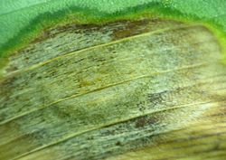
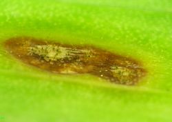
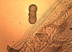
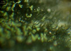
The characteristic feature of the pathogen is its infecting the leaves of the plant. On their surface there are formed elliptic, light-fulvous, with brown edge and a lighter middle part, drying spots (fig. 1, 2, 3, 4). The area of these spots is constantly growing which causes a considerable damage of the plant and even its death. In the middle part of the spots there can be formed brown clusters that consist of conidiophores being formed on the hyphae of the submersed mycelium, also of stroma and unripe perithecium (fig. 5, 6, 7, 8, 9, 10). Conidiophores are first straight, than variously flexuous, simple or branched, septate or not, often inflated at the bottom (fig. 6), dark-brown, olive, 20-200 x 6-16 µm (fig. 11, 12, 13, 14, 15). Conidia are being formed on the conidiophores (fig. 16, 17). They are often solitary (fig. 18, 19, 20, 21), sometimes are formed in the short chains, cylindrical and prolonged, acanthaceous and verrucouse, colored from soft to dark brown, 1-5 (mostly 2-3) septate, 20-60 х 15-25 µm, often slightly constricted at the septa (fig. 22, 23, 24, 25, 26, 27, 28, 29).
The fungus H. gracile grows well in culture on PDA forming up abundant aerial mycelium. The hyphae of the submersed mycelium are colored from light to dark brown, flexuous and septate with more thinner or thicker walls 3-14 µm in diameter. Stroma is black, is formed out of dense hyphae interlacement. Later typical conidiophores are being formed that can be 500 µm long, within them there are often generated endogeneous hyphae, and on the surface – conidia. Conidia are very fragile, even when slightly touched they can burst with its content flowing out (fig. 30).
- McKemy, J.M.; Morgan-Jones, G. 1990. Studies in the genus Cladosporium sensu lato. II. Concerning Heterosporium gracile, the causal organism of leaf spot disease of Iris species. Mycotaxon 39:425-440.
| 1. Iris hybrida hort. with symptoms of leaf blotch и leaf spot (Image by G. Pestsov)
|
| 2. Iris hybrida hort. leaf infected with fungus Heterosporium gracile (Image by G. Pestsov)
|
| 3. Typical symptoms of Heterosporium gracile infection. (Image by G. Pestsov)
|
| 4. Beginning of Heterosporium gracile morpholodical structures' formation on the leaf's affected tissue. (Image by G. Pestsov)
|
| 5. Leaf's necrotic tissue and formation of conidiophores' clusters. (Image by G. Pestsov)
|
| 6. Formation of conidiophores on the leaf's affected tissue. (Image by G. Pestsov)
|
| 7. Submersed mycelium of Heterosporium gracile (Image by G. Pestsov)
|
| 8. Conidiophores of Heterosporium gracile. (Image by G. Pestsov)
|
| 9. Heterosporium gracile conidiophores and conidia. (Image by G. Pestsov)
|
| 10. Вeginning of formation of Heterosporium gracile conidiophores. (Image by G. Pestsov)
|
| 11. Conidiophores and conidia of Heterosporium gracile. (Image by G. Pestsov)
|
| 12. Formation of conidiophores on the leaf's surface. (Image by G. Pestsov)
|
| 13. Сonidia, conidiophores and stromata from host. (Image by G. Pestsov)
|
| 14. Conidiophores Heterosporium gracile and stromata from host. (Image by G. Pestsov)
|
| 15. Apical growth of the Heterosporium gracile conidiophore. (Image by G. Pestsov)
|
| 16. Formation of conidia on the conidiophores. (Image by G. Pestsov)
|
| 17. Mass conidia formation. (Image by G. Pestsov)
|
| 18. Conidium of Heterosporium gracile. (Image by G. Pestsov)
|
| 19. Heterosporium gracile conidium formation on the conidiophore. (Image by G. Pestsov)
|
| 20. Formation of Heterosporium gracile conidiophores and conidia. (Image by G. Pestsov)
|
| 21. Formation of Heterosporium gracile conidium. (Image by G. Pestsov)
|
| 22. Mycelium, conidiophores and conidia of Heterosporium gracile. (Image by G. Pestsov)
|
| 23. Verruculose surface of Heterosporium gracile conidia. (Image by G. Pestsov)
|
| 24. Conidia and hyphae of endogenec Heterosporium gracile mycelium. (Image by G. Pestsov)
|
| 25. Acanthaceously verrucate conidia surface of Heterosporium gracile. (Image by G. Pestsov)
|
| 26. Сonidium and conidiophore of Heterosporium gracile. (Image by G. Pestsov)
|
| 27. Heterosporium gracile conidia colored with fuchsin. (Image by G. Pestsov)
|
| 28. Septate conidia with constrictions of Heterosporium gracile. (Image by G. Pestsov)
|
| 29. Septa of Heterosporium gracile conidia. (Image by G. Pestsov)
|
| 30. Fractured conidium of Heterosporium gracile. (Image by G. Pestsov)
|
|
| All images by G. Pestsov. |


