Diptacus berberinus
| Notice: | This page is derived from the original publication listed below, whose author(s) should always be credited. Further contributors may edit and improve the content of this page and, consequently, need to be credited as well (see page history). Any assessment of factual correctness requires a careful review of the original article as well as of subsequent contributions.
If you are uncertain whether your planned contribution is correct or not, we suggest that you use the associated discussion page instead of editing the page directly. This page should be cited as follows (rationale):
Citation formats to copy and paste
BibTeX: @article{Li2012ZooKeys196, RIS/ Endnote: TY - JOUR Wikipedia/ Citizendium: <ref name="Li2012ZooKeys196">{{Citation See also the citation download page at the journal. |
Ordo: Trombidiformes
Familia: Eriophyidae
Genus: Diptacus
Name
Diptacus berberinus Li & Xue & Hong, 2012 sp. n. – Wikispecies link – ZooBank link – Pensoft Profile
Description
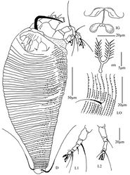
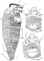
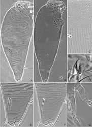
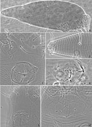
Female. (n = 9) Body fusiform, light yellow, 283 (280–360), 110 (102–110) wide, 115 (114–115) thick. Gnathosoma 26 (25–27), projecting downwards, pedipalp coxal seta (ep) 6 (5–6), dorsal pedipalp genual seta (d) 12 (11–12), cheliceral stylets 65 (65–66). Prodorsal shield 40 (40–46), 80 (77–80) wide, with wide and broad frontal lobe, 7 (7–8); median, admedian and submedian lines present, admedian lines connected at the basal 1/3 and 2/3 of prodorsal shield, forming 3 cells on each side, submedian lines connected with the median and admedian at the basal 2/3 of prodorsal shield, forming the cell-like pattern at anterior shield margin. Scapular tubercles ahead of rear shield margin, 3 (2–3), 30 (27–30) apart, scapular setae (sc) 5 (4–5), projecting centrad to forward. Coxigenital region with 13 (13–15) annuli, with triangular microtubercles. Coxisternal plate I with granules, coxisternal plate II smooth, anterolateral setae on coxisternum I (1b) 20 (18–20), 17 (17–18) apart, proximal setae on coxisternum I (1a) 43 (43–45), 19 (17–19) apart, proximal setae on coxisternum II (2a) 70 (70–80), 49 (43–56) apart, tubercles 1b and 1a 12 (12–13) apart, tubercles 1a and 2a 17 (14–17) apart. Prosternal apodeme separated, 5 (5–6). Leg I 65 (60–65), femur 20 (20–22), basiventral femoral seta (bv) absent; genu 8 (8–9), antaxial genual seta (l") 48 (48–52); tibia 18 (17–19), paraxial tibial seta (l’) 11 (10–11), located at 1/2 from dorsal base; tarsus 11 (11–12), seta ft’ 30 (29–30), seta ft" 40 (37–40), seta u’ 7 (6–7); tarsal empodium (em) 11 (10–11), divided, 7-rayed on each side, tarsal solenidion (ω) 10 (10–14), knobbed. Leg II 55 (54–55), femur 20 (19–20), basiventral femoral seta (bv) absent; genu 8 (7–8), antaxial genual seta (l") 18 (15–18); tibia 17 (16–17); tarsus 11 (10–11), seta ft’ 11 (10–11), seta ft" 44 (44–50), seta u’ 8 (7–8); tarsal empodium (em) 11 (10–11), divided, 7-rayed on each side, tarsal solenidion (ω) 10 (10–11), knobbed. Opisthosoma dorsally with 59 (54–62) annuli, smooth, ventrally with 106 (101–106) annuli, with triangular microtubercles. Setae c2 115 (110–115) on ventral annulus 18 (18–20), 74 (74–75) apart; setae d 100 (100–120) on ventral annulus 40 (37–40), 56 (49–56) apart; setae e 60 (55–60) on ventral annulus 65 (60–65), 31 (29–35) apart; setae f 65 (60–70) on ventral annulus 93 (87–93), 35 (35–37) apart. Setae h1 2 (1–2), h2 103 (95–165). Female genitalia 35 (31–40), 35 (34–42) wide, coverflap with short lines on base, and 4 longitudinal ridges in 2 ranks, 1 ridge near the base and 3 ridges at distal margin, setae 3a 12 (11–15), 24 (24–25) apart.
Male. (n = 1) Body fusiform, light yellow, 269, 86 wide. Gnathosoma 60, projecting downwards, pedipalp coxal seta (ep) 5, dorsal pedipalp genual seta (d) 11, cheliceral stylets 65. Prodorsal shield has the same design as female, 38, 71 wide, with wide and broad frontal lobe, 7. Scapular tubercles ahead of rear shield margin, 3, 27 apart, scapular setae (sc) 4, projecting centrad to forward. Coxigenital region with 14 annuli, with triangular microtubercles. Coxisternal plate I with granules, coxisternal plate II smooth, anterolateral setae on coxisternum I (1b) 22, 16 apart, proximal setae on coxisternum I (1a) 36, 16 apart, proximal setae on coxisternum II (2a) 60, 41 apart, tubercles 1b and 1a 11 apart, tubercles 1a and 2a 12 apart. Prosternal apodeme separated, 6. Leg I 46, femur 17, basiventral femoral seta (bv) absent; genu 7, antaxial genual seta (l") 46; tibia 13, paraxial tibial seta (l’) 10, located at 1/2 from dorsal base; tarsus 8, seta ft’ 30, seta ft" 37, seta u’ 6; tarsal empodium (em) 10, divided, 7-rayed on each side, tarsal solenidion (ω) 10, knobbed. Leg II 38, femur 17, basiventral femoral seta (bv) absent; genu 7, antaxial genual seta (l") 15; tibia 13; tarsus 6, seta ft’ 10, seta ft" 38, seta u’ 7; tarsal empodium (em) 10, divided, 7-rayed on each side, tarsal solenidion (ω) 11, knobbed. Opisthosoma dorsally with 56 annuli, smooth, ventrally with 83 annuli, with triangular microtubercles. Setae c2 95 on ventral annulus 15, 63 apart; setae d 100 on ventral annulus 30, 49 apart; setae e 55 on ventral annulus 46, 27 apart; setae f 60 on ventral annulus 71, 25 apart. Setae h1 2, h2 120. Male genitalia 23, 30 wide, setae 3a 10, 23 apart.
Type material
Holotype, female (slide number 783, marked Holotype), from Berberis amurensis Rupr.(Berberidaceae), Mengda Natural Reserve, Xunhua County, Qinghai Province, P. R. China, 35°47'38"N, 102°40'40"E, elevation 2523m, 19 July 2007, coll. Xiao-Feng Xue. Paratypes, 8 females and 1 male (slide number 783), with the same data as holotype.
Relation to host
Vagrant on leaf lower surface. No damage to the host was observed.
Etymology
The specific designation berberinus is from the generic name of host plant, Berberis; masculine in gender.
Differential diagnosis
This species is similar to Diptacus maddenis Song, Xue & Hong, 2007a, but can be differentiated from the latter by opisthosomal dorsal annuli smooth (opisthosomal dorsal annuli with elongated microtubercles in Diptacus maddenis), female genital coverflap with short lines at the base (genital coverflap with granules in Diptacus maddenis), tarsal empodium 7-rayed (4-rayed in Diptacus maddenis).
Original Description
- Li, H; Xue, X; Hong, X; 2012: Eriophyoid mites from Qinghai Province, northwestern China with descriptions of nine new species (Acari, Eriophyoidea) ZooKeys, 196: 47-107. doi
Images
|


