Sclerotinia Disease of Carrots
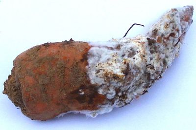
Figure 1. Carrot infected with Sclerotinia disease. Image by G. Pestsov.
When storing carrots one usually confronts a problem – how to prevent the roots from being infected with watery soft rot. The disease was given this name because of the specific appearance of the affected root that is covered with the white mycelium consisting of a great number of the interlaced hyphae (Fig. 1). This disease is also called Sclerotinia disease (after the name of the pathogen - Sclerotinia sclerotiorum (Lib.) dBy [syn.: Whetzelinia sclerotiorum (Lib.) de Bary]).
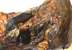
The mycelium of the fungi shoots fast and under auspicious conditions can affect any number of carrots put in storage (Fig. 2). At that the tissue of the roots softens, but doesn’t lose its colour. This happens under the action of the ferment pectinase that is synthesized by the fungi in great quantities.
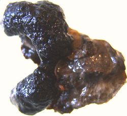
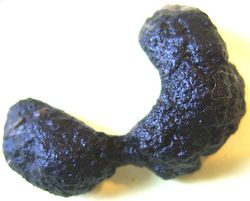
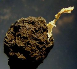
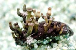
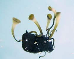
With time on the carrot’s affected tissue there appear large, with the size of 1-3 cm, sclerotia, consisting of the tightly interlaced hyphae. In the beginning they are white, then are getting darker, finally turning black (fig. 3, 4, 5). Sclerotia help the phytopathogen to stand adverse conditions. The period of the dormancy ends in spring, the sclerotia grow out forming up the apothecia (fig. 6, 7, 8). The apothecia always grow in the direction of light, which means they have positive phototropism.
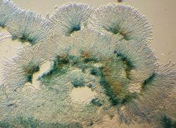
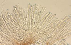
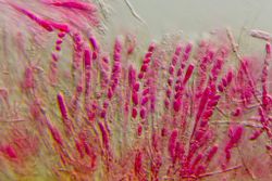
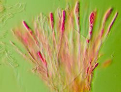
First, there appears a stem that then becomes longer widening at the end and forming a funnel up to 9 mm in diameter (fig. 8), in which asci and ascospores are being formed (fig. 9, 10, 11, 12). Iodine colors the asci apices into color blue (fig. 13, 14). The ascospores are thrown out of the asci with great force of the osmotic pressure and then are spread around by the wind. On getting into the favorable substratum the spores grow out into the vegetative primary mycelium. If the spore, for instance, fell into the pistil stigma of the plant host, it would easily get into the inner parts of the flower, and then in the vegetative parts of the plant. The primary inoculation has thus occurred, the disease development may be latent during the vegetation, but will definitely show when the roots are stored.
| Image gallery showing Sclerotinia on carrots |
| Figure 2. Stored carrot showing severe watery soft rot symptoms.
|
| Figure 3. Mature sclerotia that were formed in the macerative tissue of the carrot’s root.
|
| Figure 4. Mature sclerotium.
|
| Figure 5. Mature sclerotium.
|
| Figure 6. Defective apothecia formation.
|
| Figure 7. Apothecia formation.
|
| Figure 8. Sclerotinia sclerotiorum apothecia formation.
|
| Figure 9. Asci formation in apothecia.
|
| Figure 10. Asci with ascospores and paraphysis.
|
| Figure 11. Asci with ascospores and paraphysis.
|
| Figure 12. Asci with ascospores and paraphysis.
|
| Figure 13. Asci with ascospores colored by iodine.
|
| Figure 14. Asci with ascospores colored by iodine.
|
|
| All images by Georgy Pestsov. |
(This page was moved here from http://phytopathology.net/Portal/Sclerotinia_disease )


