Difference between revisions of "Cycloporus papillosus"
m (Imported from ZooKeys) |
m (1 revision) |
(No difference)
| |
Latest revision as of 15:13, 22 April 2014
| Notice: | This page is derived from the original publication listed below, whose author(s) should always be credited. Further contributors may edit and improve the content of this page and, consequently, need to be credited as well (see page history). Any assessment of factual correctness requires a careful review of the original article as well as of subsequent contributions.
If you are uncertain whether your planned contribution is correct or not, we suggest that you use the associated discussion page instead of editing the page directly. This page should be cited as follows (rationale):
Citation formats to copy and paste
BibTeX: @article{Noreña2014ZooKeys404, RIS/ Endnote: TY - JOUR Wikipedia/ Citizendium: <ref name="Noreña2014ZooKeys404">{{Citation See also the citation download page at the journal. |
Ordo: Polycladida
Familia: Euryleptidae
Genus: Cycloporus
Name
Cycloporus papillosus (Sars, 1878) Lang, 1884 – Wikispecies link – Pensoft Profile
Material examined
Four individuals captured during summer, autumn and winter between 2010 and 2012 (11/10/2010; 03/08/2011; 27/01/2011; 09/01/2012). Voucher: one specimen sectioned sagittally, stained with azan and deposited in the Invertebrate Collections of the MNCN; Cat. Nr: MNCN 4.01/573 to 4.01/599 (27 slides). Further material: one specimen cat. Nr. MNCN 4.01/600 to 4.01/625 (26 slides).
Description
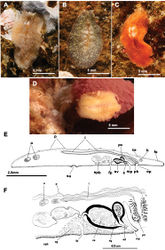
Elongated worms 11 mm long and 6 mm wide. Body shape elongated with light undulating margins and rounded anterior and posterior ends. Dorsal surface with numerous papillae. Colouration orange, yellowish orange or translucent grey with white patches at the mid-dorsal line (Figure 1A, B, C, D). Ventral side smooth and pale. Short inconspicuous marginal tentacles. Sucker located approximately in the middle of the body (Figure 1E). Tentacular eyes scattered over dorsal margin of tentacles (Figure 1A), cerebral eyes in two elongated, anteriorly anastomosing clusters. Plicate cylindrical or tubular pharynx near anterior end, frontally oriented; oral pore posterior to brain. Male and female genital pores clearly separated and posterior to pharynx (Figure 1E). Male copulatory apparatus located posterior to male pore and oriented mainly dorso -ventrally, but also directed frontally (Figure 1E, F). Male system consists of a short, armed (stylet) penis papilla, a true prostatic vesicle with a smooth glandular epithelium, and a well developed, muscularized seminal vesicle. Prostatic vesicle opens directly into penis papilla, and seminal vesicle empties directly into distal end of prostatic vesicle. Vasa deferentia, sometimes very dilated, open proximally through a common duct (common vas deferens) into seminal vesicle.
Inconspicuous female system lies posterior to the male pore and is characterized by a short female atrium, female duct (vagina), and characteristic uterine vesicles. Abundant cement glands are located around female pore and distal part of vagina.
Remarks on biology
Cycloporus papillosus is a natural predator of Botrylloides violaceus Oka, 1927 (Ascidiacea), which is a clear example of an invasive species. Botrylloides violaceus grows on all types of substrates, including other living animals such as mussels, small sea cucumbers or other ascidians, covering them completely and killing them. Botrylloides violaceus has completely replaced Botrylloides leachii, the autochthonous ascidian in this area. Both species of ascidians compete for the same substrate. Cycloporus papillosus preys on Botrylloides violaceus and places its egg plates (Fig. 1D) in the folds of this species (in the area where new zooids grow and extend the colony) or under the unattached colony, thereby ensuring larval protection and the availability of food after hatching.
Distribution
In Galicia, three specimens of Cycloporus papillosus were captured from mussels collected on Botrylloides violaceus on the docks of the Yacht Club Ribeira (Ria de Arosa, Galicia, Spain). Depth varied between 0.5 and 1 metres (42°33.7770N, 008°59.2970W; 42°33.7850N, 008°59.3140W; 42°33.7930N, 008°59.3290W). Another specimen (Figure 1B) was collected on a colony of Botryllus schlosseri (Ascidiacea) growing on a rock of the island of Rua (Ria de Arousa, Galicia, Spain), at a depth of 14 metres (42°32.9650N, 008°56.4590W).
This is the southernmost European record for Cycloporus papillosus, and the first for the Atlantic coast of the Iberian Peninsula. Other localities from where this polyclad has been reported are: Bergen, Norway (Jensen 1878[1]); Rovigno, Croatia (Vàtova 1928[2]); Susaki near Simoda, Japan (Kato 1937[3]); Porto Praia, Cape Verde (Laidlaw 1906[4]); Plymouth, United Kingdom (Gamble 1893[5]).
Taxon Treatment
- Noreña, C; Marquina, D; Perez, J; Almon, B; 2014: First records of Cotylea (Polycladida, Platyhelminthes) for the Atlantic coast of the Iberian Peninsula ZooKeys, 404: 1-22. doi
Other References
- ↑ Jensen O (1878) Turbellaria ad litora Norvegiae occidentalia. Turbellarier ved Norges Vestkyst. J W Eided Bogtrykkeri, Bergen, 97 pp.
- ↑ Vàtova A (1928) Compendis della flora e fauna del Mare Adriatico presso Rovigno con la distribuzione geografica delle species bentoniche. Memoria - Comitato talassografico italiano 143: 154-174.
- ↑ Kato K (1937) Polyclads collected in Idu, Japan. Japanese Journal of Zoology 7: 211-232.
- ↑ Laidlaw F (1906) On the marine fauna of the Cape Verde Islands, from collections made in 1904 by Mr C. Crossland. The polyclad Turbellaria. Proceedings of the Zoological Society of London, 1906, 705–719.
- ↑ Gamble F (1893) The Turbellaria of Plymouth Sound and the neighbourhood. Journal of the Marine Biological Associations (N S) 3(1): 18, 30–47.
Images
|
