Difference between revisions of "Heterosporium gracile"
(removing the history to show normal page) |
|||
| (3 intermediate revisions by the same user not shown) | |||
| Line 1: | Line 1: | ||
Many species of the genus ''Heterosporium'' Klotzsch. can lead a saprophytic lifesyle, but more often they parasitize on the wild and cultivated plants from different botanic families. They affect plants of the genera ''Allium, Avena, Hordeum, Secale, Beta, Sorghum, Brassica, Zea, Robinia, Dianthus, Lychnis, Saponaria, Freesia, Hemerocallis, Sambucus, Galtonia, Narcissus, Gladiolus, Iris''. The fungus ''Heterosporium gracile'' Sacc.* is a causal agent of leaf blotch or leaf spot of different species and cultivars ''Iris'' L. both in Europe and America. | Many species of the genus ''Heterosporium'' Klotzsch. can lead a saprophytic lifesyle, but more often they parasitize on the wild and cultivated plants from different botanic families. They affect plants of the genera ''Allium, Avena, Hordeum, Secale, Beta, Sorghum, Brassica, Zea, Robinia, Dianthus, Lychnis, Saponaria, Freesia, Hemerocallis, Sambucus, Galtonia, Narcissus, Gladiolus, Iris''. The fungus ''Heterosporium gracile'' Sacc.* is a causal agent of leaf blotch or leaf spot of different species and cultivars ''Iris'' L. both in Europe and America. | ||
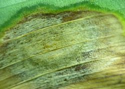
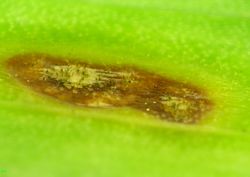
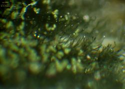
| − | The characteristic feature of the pathogen is its infecting the leaves of the plant. On their surface there are formed elliptic, light-fulvous, with brown edge and a lighter middle part, drying spots (fig. 1, 2, 3, 4). The area of these spots is constantly growing which causes a considerable damage of the plant and even its death. In the middle part of the spots there can be formed brown clusters that consist of conidiophores being formed on the hyphae of the submersed mycelium, also of stroma and unripe perithecium (fig. 5, 6, 7, 8, 9, 10). Conidiophores are first straight, than variously flexuous, simple or branched, septate or not, often inflated at the bottom (fig. 6), dark-brown, olive, 20-200 x 6-16 µm (fig. 11, 12, 13, 14, 15). Conidia are being formed on the conidiophores (fig. 16, 17). They are often solitary (fig. 18, 19, 20, 21), sometimes are formed in the short chains, cylindrical and prolonged, acanthaceous and verrucouse, colored from soft to dark brown, 1-5 (mostly 2-3) septate, 20-60 х 15-25 µm, often slightly constricted at the septa (fig. 22, 23, 24, 25, 26, 27, 28, 29). | + | The characteristic feature of the pathogen is its infecting the leaves of the plant. On their surface there are formed elliptic, light-fulvous, with brown edge and a lighter middle part, drying spots (fig. 1, 2, 3, 4). The area of these spots is constantly growing which causes a considerable damage of the plant and even its death. In the middle part of the spots there can be formed brown clusters that consist of conidiophores being formed on the hyphae of the submersed mycelium, also of stroma and unripe perithecium (fig. 5, 6, 7, 8, 9, 10). Conidiophores are first straight, than variously flexuous, simple or branched, septate or not, often inflated at the bottom (fig. 6), dark-brown, olive, 20-200 x 6-16 µm (fig. 11, 12, 13, 14, 15). Conidia are being formed on the conidiophores (fig. 16, 17). They are often solitary (fig. 18, 19, 20, 21), sometimes are formed in the short chains, cylindrical and prolonged, acanthaceous and verrucouse, colored from soft to dark brown, 1-5 (mostly 2-3) septate, 20-60 х 15-25 µm, often slightly constricted at the septa (fig. 22, 23, 24, 25, 26, 27, 28, 29). |
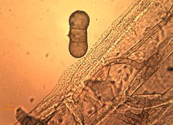
The fungus ''H. gracile'' grows well in culture on PDA forming up abundant aerial mycelium. The hyphae of the submersed mycelium are colored from light to dark brown, flexuous and septate with more thinner or thicker walls 3-14 µm in diameter. Stroma is black, is formed out of dense hyphae interlacement. Later typical conidiophores are being formed that can be 500 µm long, within them there are often generated endogeneous hyphae, and on the surface – conidia. Conidia are very fragile, even when slightly touched they can burst with its content flowing out (fig. 30). | The fungus ''H. gracile'' grows well in culture on PDA forming up abundant aerial mycelium. The hyphae of the submersed mycelium are colored from light to dark brown, flexuous and septate with more thinner or thicker walls 3-14 µm in diameter. Stroma is black, is formed out of dense hyphae interlacement. Later typical conidiophores are being formed that can be 500 µm long, within them there are often generated endogeneous hyphae, and on the surface – conidia. Conidia are very fragile, even when slightly touched they can burst with its content flowing out (fig. 30). | ||
| Line 9: | Line 9: | ||
{{Gallery | {{Gallery | ||
| − | | footer= All images by | + | | footer= All images by Georgy Pestsov. |
| width=250 | | width=250 | ||
| lines=3 | | lines=3 | ||
| − | | File:Iris hybrida hort. with symptoms of leaf blotch и leaf spot.JPG |1. ''Iris hybrida'' hort. with symptoms of leaf blotch и leaf spot (Image by G. Pestsov) | + | | File:Iris hybrida hort. with symptoms of leaf blotch и leaf spot.JPG |1. ''Iris hybrida'' hort. with symptoms of leaf blotch и leaf spot. (Image by G. Pestsov) |
| − | | File: Iris hybrida hort. leaf infected with fungus Heterosporium gracile.JPG |2. ''Iris hybrida'' hort. leaf infected with fungus ''Heterosporium gracile'' (Image by G. Pestsov) | + | | File: Iris hybrida hort. leaf infected with fungus Heterosporium gracile.JPG |2. ''Iris hybrida'' hort. leaf infected with fungus ''Heterosporium gracile''. (Image by G. Pestsov) |
| File:Typical symptoms of Heterosporium gracile infection.JPG |3. Typical symptoms of ''Heterosporium gracile'' infection. (Image by G. Pestsov) | | File:Typical symptoms of Heterosporium gracile infection.JPG |3. Typical symptoms of ''Heterosporium gracile'' infection. (Image by G. Pestsov) | ||
| File:Beginning of Heterosporium gracile morpholodical structures formation on the leafs affected tissue.jpg |4. Beginning of ''Heterosporium gracile'' morpholodical structures' formation on the leaf's affected tissue. (Image by G. Pestsov) | | File:Beginning of Heterosporium gracile morpholodical structures formation on the leafs affected tissue.jpg |4. Beginning of ''Heterosporium gracile'' morpholodical structures' formation on the leaf's affected tissue. (Image by G. Pestsov) | ||
| File:5. Leafs necrotic tissue and formation of conidiophores clusters.jpg |5. Leaf's necrotic tissue and formation of conidiophores' clusters. (Image by G. Pestsov) | | File:5. Leafs necrotic tissue and formation of conidiophores clusters.jpg |5. Leaf's necrotic tissue and formation of conidiophores' clusters. (Image by G. Pestsov) | ||
| File:Formation of conidiophores on the leafs affected tissue.jpg |6. Formation of conidiophores on the leaf's affected tissue. (Image by G. Pestsov) | | File:Formation of conidiophores on the leafs affected tissue.jpg |6. Formation of conidiophores on the leaf's affected tissue. (Image by G. Pestsov) | ||
| − | | File:Submersed mycelium of Heterosporium gracile.jpg |7. Submersed mycelium of ''Heterosporium gracile'' (Image by G. Pestsov) | + | | File:Submersed mycelium of Heterosporium gracile.jpg |7. Submersed mycelium of ''Heterosporium gracile''. (Image by G. Pestsov) |
| File:Conidiophores of Heterosporium gracile.jpg |8. Conidiophores of ''Heterosporium gracile''. (Image by G. Pestsov) | | File:Conidiophores of Heterosporium gracile.jpg |8. Conidiophores of ''Heterosporium gracile''. (Image by G. Pestsov) | ||
| File:Heterosporium gracile conidiophores and conidia.JPG |9. ''Heterosporium gracile'' conidiophores and conidia. (Image by G. Pestsov) | | File:Heterosporium gracile conidiophores and conidia.JPG |9. ''Heterosporium gracile'' conidiophores and conidia. (Image by G. Pestsov) | ||
| Line 41: | Line 41: | ||
| File:Septate conidia with constrictions of Heterosporium gracile.JPG |28. Septate conidia with constrictions of ''Heterosporium gracile''. (Image by G. Pestsov) | | File:Septate conidia with constrictions of Heterosporium gracile.JPG |28. Septate conidia with constrictions of ''Heterosporium gracile''. (Image by G. Pestsov) | ||
| File:Septa of Heterosporium gracile conidia.JPG |29. Septa of ''Heterosporium gracile'' conidia. (Image by G. Pestsov) | | File:Septa of Heterosporium gracile conidia.JPG |29. Septa of ''Heterosporium gracile'' conidia. (Image by G. Pestsov) | ||
| − | | File:Fractured conidium of Heterosporium gracile.JPG |30. Fractured conidium of ''Heterosporium gracile''. (Image by G. Pestsov) | + | | File:Fractured conidium of Heterosporium gracile.JPG |30. Fractured conidium of ''Heterosporium gracile''. (Image by G.V. Pestsov) |
}} | }} | ||
(This page was moved here from http://phytopathology.net/Portal/Heterosporium_gracile ) | (This page was moved here from http://phytopathology.net/Portal/Heterosporium_gracile ) | ||
Latest revision as of 20:48, 21 March 2022
Many species of the genus Heterosporium Klotzsch. can lead a saprophytic lifesyle, but more often they parasitize on the wild and cultivated plants from different botanic families. They affect plants of the genera Allium, Avena, Hordeum, Secale, Beta, Sorghum, Brassica, Zea, Robinia, Dianthus, Lychnis, Saponaria, Freesia, Hemerocallis, Sambucus, Galtonia, Narcissus, Gladiolus, Iris. The fungus Heterosporium gracile Sacc.* is a causal agent of leaf blotch or leaf spot of different species and cultivars Iris L. both in Europe and America.
The characteristic feature of the pathogen is its infecting the leaves of the plant. On their surface there are formed elliptic, light-fulvous, with brown edge and a lighter middle part, drying spots (fig. 1, 2, 3, 4). The area of these spots is constantly growing which causes a considerable damage of the plant and even its death. In the middle part of the spots there can be formed brown clusters that consist of conidiophores being formed on the hyphae of the submersed mycelium, also of stroma and unripe perithecium (fig. 5, 6, 7, 8, 9, 10). Conidiophores are first straight, than variously flexuous, simple or branched, septate or not, often inflated at the bottom (fig. 6), dark-brown, olive, 20-200 x 6-16 µm (fig. 11, 12, 13, 14, 15). Conidia are being formed on the conidiophores (fig. 16, 17). They are often solitary (fig. 18, 19, 20, 21), sometimes are formed in the short chains, cylindrical and prolonged, acanthaceous and verrucouse, colored from soft to dark brown, 1-5 (mostly 2-3) septate, 20-60 х 15-25 µm, often slightly constricted at the septa (fig. 22, 23, 24, 25, 26, 27, 28, 29).
The fungus H. gracile grows well in culture on PDA forming up abundant aerial mycelium. The hyphae of the submersed mycelium are colored from light to dark brown, flexuous and septate with more thinner or thicker walls 3-14 µm in diameter. Stroma is black, is formed out of dense hyphae interlacement. Later typical conidiophores are being formed that can be 500 µm long, within them there are often generated endogeneous hyphae, and on the surface – conidia. Conidia are very fragile, even when slightly touched they can burst with its content flowing out (fig. 30).
- McKemy, J.M.; Morgan-Jones, G. 1990. Studies in the genus Cladosporium sensu lato. II. Concerning Heterosporium gracile, the causal organism of leaf spot disease of Iris species. Mycotaxon 39:425-440.
| ||||||||||||||||||||||||||||||||||||||||||||||||||||||||||||
| All images by Georgy Pestsov. | ||||||||||||||||||||||||||||||||||||||||||||||||||||||||||||
(This page was moved here from http://phytopathology.net/Portal/Heterosporium_gracile )


