Difference between revisions of "Blue Moulds of Oranges"
(The article is by Georgy Pestsov; Gregor Hagedorn only moved the content to a new wiki after the original had to be given up) |
|||
| (3 intermediate revisions by the same user not shown) | |||
| Line 7: | Line 7: | ||
In certain places, often at places of the peel injury, the peel is growing softer and acquires a watery consistence that is very distinct from the healthy one (fig. 3, 4). Tissue dissociation results from the fungi action. Gradually they insinuate in the inside of the fruit, changing its consistence, composition and coloring of the pulp. | In certain places, often at places of the peel injury, the peel is growing softer and acquires a watery consistence that is very distinct from the healthy one (fig. 3, 4). Tissue dissociation results from the fungi action. Gradually they insinuate in the inside of the fruit, changing its consistence, composition and coloring of the pulp. | ||
| − | Inside the fruit the fungi moves in the intercellular space and actively produces conidia inside the fruit (fig. 5). If an orange slice is kept under room temperature for 24 hours, mass sporification takes place in places already previously infected by the mycelium invisible to the naked eye (fig. 6). Sporulation occurs on asymmetrical conidiophores, bearing conidiogeneous cell producing billions of the rounded conidia in chains (fig. 7, 8, 9, 10, 11, 12). The conidia are dispersed and may damage other products. | + | Inside the fruit the fungi moves in the intercellular space and actively produces conidia inside the fruit (fig. 5). If an orange slice is kept under room temperature for 24 hours, mass sporification takes place in places already previously infected by the mycelium invisible to the naked eye (fig. 6). Sporulation occurs on asymmetrical conidiophores, bearing conidiogeneous cell producing billions of the rounded conidia in chains (fig. 7, 8, 9, 10, 11, 12). The conidia are dispersed and may damage other products. |
People, especially children, can suffer allergic reactions when inhaling or consuming the conidia. | People, especially children, can suffer allergic reactions when inhaling or consuming the conidia. | ||
| Line 14: | Line 14: | ||
{{Gallery | {{Gallery | ||
| − | | footer= All images by | + | | footer= All images by Georgy Pestsov. |
| width=250 | | width=250 | ||
| lines=4 | | lines=4 | ||
| Line 27: | Line 27: | ||
| File: Formation of conidia on the conidiophores with assymetrical penicillus.jpg | Figure 8.Formation of conidia on the conidiophores with assymetrical penicillus.jpg (Image by G. Pestsov) | | File: Formation of conidia on the conidiophores with assymetrical penicillus.jpg | Figure 8.Formation of conidia on the conidiophores with assymetrical penicillus.jpg (Image by G. Pestsov) | ||
| File:Blue Mould of Oranges - Conidiophores.jpg | Figure 9.Conidiophores of the fungus ''Penicillium italicum''.jpg (Image by G. Pestsov) | | File:Blue Mould of Oranges - Conidiophores.jpg | Figure 9.Conidiophores of the fungus ''Penicillium italicum''.jpg (Image by G. Pestsov) | ||
| − | | File: Conidia formation on sterigma.jpg | Figure 10.Conidia formation on sterigma.jpg (Image by G. Pestsov) | + | | File: Conidia formation on sterigma.jpg | Figure 10. Conidia formation on sterigma.jpg (Image by G. Pestsov) |
| − | | File: 8. Formation of the conidiophores.jpg | Figure 11. Formation of the conidiophores by the fungus ''Penicillium italicum''.jpg (Image by G. Pestsov) | + | | File: 8. Formation of the conidiophores.jpg | Figure 11. Formation of the conidiophores by the fungus ''Penicillium italicum''.jpg (Image by G.V. Pestsov) |
| File:Isolated conidiophore.jpg | Figure 12.Isolated conidiophore.jpg (Image by G. Pestsov) | | File:Isolated conidiophore.jpg | Figure 12.Isolated conidiophore.jpg (Image by G. Pestsov) | ||
}} | }} | ||
Latest revision as of 20:39, 21 March 2022
By Georgy Pestsov (page moved here from http://phytopathology.net/Portal/Blue_Mould_of_Oranges )
Due to their superb taste and nutritional value, oranges figure prominently in the world’s production of citrus plants. Their refreshing taste is determined by a combination of sugar, organic acids, pectines, and vitamin C. However, when gathered, transported or stored, oranges can be infected by fungal diseases. When preserved under low temperatures, the disease symptoms usually don’t show. As soon as conditions grow favorable, phytopathogens reproduce intensively.
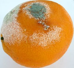
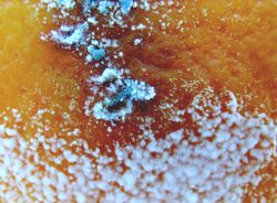
Infection of oranges by the Blue Mould (pathogen Penicillium italicum Wehmer) often occurs through the fruit’s peel lesion. Mycelium of the fungi start growing inside of the peel, and under room temperature quickly occupies new space (fig. 1). Initially small white colonies appear on the surface, rapidly growing larger. Within several hours they become grayish-green and in 24 hours grayish-blue. The color change is a result of the spore formation. The older the colony, the more spores are being produced and the more intensively it is thus colored (fig. 2).
In certain places, often at places of the peel injury, the peel is growing softer and acquires a watery consistence that is very distinct from the healthy one (fig. 3, 4). Tissue dissociation results from the fungi action. Gradually they insinuate in the inside of the fruit, changing its consistence, composition and coloring of the pulp.
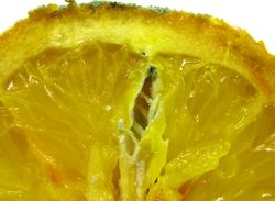
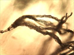
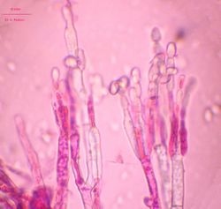
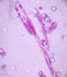
Inside the fruit the fungi moves in the intercellular space and actively produces conidia inside the fruit (fig. 5). If an orange slice is kept under room temperature for 24 hours, mass sporification takes place in places already previously infected by the mycelium invisible to the naked eye (fig. 6). Sporulation occurs on asymmetrical conidiophores, bearing conidiogeneous cell producing billions of the rounded conidia in chains (fig. 7, 8, 9, 10, 11, 12). The conidia are dispersed and may damage other products.
People, especially children, can suffer allergic reactions when inhaling or consuming the conidia.
The following images illustrate the development of blue mould on oranges.
| ||||||||||||||||||||||||
| All images by Georgy Pestsov. | ||||||||||||||||||||||||
