Difference between revisions of "Abrictosaurus consors"
m (Imported from ZooKeys) |
m (1 revision) |
(No difference)
| |
Revision as of 14:03, 3 October 2012
Contents
- 1 Taxonavigation
- 2 Name
- 3 Holotype
- 4 Type locality
- 5 Horizon
- 6 Revised diagnosis
- 7 Comments
- 8 Description
- 9 Premaxilla
- 10 Maxilla, lacrimal, postorbital, and palpebral
- 11 Quadratojugal and quadrate
- 12 Lower jaw
- 13 Premaxillary teeth
- 14 Maxillary teeth
- 15 Dentary teeth
- 16 Skull reconstruction
- 17 Axial skeleton
- 18 Pectoral girdle
- 19 Forelimb
- 20 Pelvic girdle
- 21 Hindlimb
- 22 Taxon Treatment
- 23 Other References
- 24 Images
| Notice: | This page is derived from the original publication listed below, whose author(s) should always be credited. Further contributors may edit and improve the content of this page and, consequently, need to be credited as well (see page history). Any assessment of factual correctness requires a careful review of the original article as well as of subsequent contributions.
If you are uncertain whether your planned contribution is correct or not, we suggest that you use the associated discussion page instead of editing the page directly. This page should be cited as follows (rationale):
Citation formats to copy and paste
BibTeX: @article{Sereno2012ZooKeys226, RIS/ Endnote: TY - JOUR Wikipedia/ Citizendium: <ref name="Sereno2012ZooKeys226">{{Citation See also the citation download page at the journal. |
Ordo: Ornithischia
Familia: Heterodontosauridae
Genus: Abrictosaurus
Name
Abrictosaurus consors (Thulborn, 1974) – Wikispecies link – Pensoft Profile
- Abrictosaurus consors (Thulborn, 1974) – Thulborn (1974[1], Figs 2–4); Hopson (1975[2], fig. 3c, d); Galton (1986[3], fig. 16.6m); Smith (1997[4], fig. 3c, d); Norman et al. (2011[5], fig. 39A, B)
Holotype
NHMUK RU B54, ventral portion of a skull and articulated skeleton lacking the mid and distal caudal vertebrae, right coracoid, left carpus, portions of the left manus, and portions of the right hindlimb (Table 1). The author and other researchers have examined fragmentary postcranial material of a second individual catalogued with the holotypic specimen before their transfer to the NHMUK collection. At present there is no evidence that this material pertains to Abrictosaurus consors (Norman et al. 2011[5]: 236–237).
Type locality
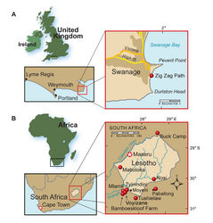
Stream-side exposure by the town Nosi (= “Noosi”), 8.2 km east of Whitehill, southern Lesotho; S30°03', E28°32' (Thulborn 1974[1]; Kitching and Raath 1984[6]) (Fig. 1B).
Horizon
Top unit (dull red sandstone) of the Upper Elliot Formation; Lower Jurassic, Hettangian, ca. 202-197 Ma (Thulborn 1974[1]; Knoll 2005[7]; Gradstein and Ogg 2009[8]).
Revised diagnosis
Heterodontosaurid ornithischian characterized by the following autapomorphies: (1) premaxillary tooth 2 and 3 with tall, subcylindrical crowns, the latter possibly representing a reduced caniniform tooth; (2) dentary tooth 2 with subcylindrical crown, possibly representing a reduced caniniform tooth; (3) maxillary and dentary teeth with flat lateral and medial crown surfaces lacking discrete marginal or median ridges; and (4) maxillary crowns in the middle of the tooth row have deep parallel-sided crowns that do not expand in mesiodistal width toward their apex. Unlike other heterodontosaurids, there is no dentary or premaxillary caniniform teeth, although both the dentary and premaxillary have teeth with subcylindrical crowns that may be reduced caniniform teeth.
Comments
A small set of autapomorphies diagnose this genus and species, which otherwise closely resembles Heterodontosaurus tucki. The lengthy initial diagnosis for Abrictosaurus consors (as Lycorhinus consors; Thulborn 1974[1]: 153–154) amounted to an abbreviated description. The only uniquely derived feature in the diagnosis (two premaxillary teeth) is erroneous, as the base of a third premaxillary tooth is present (Fig. 31). The revised diagnosis in Hopson (1975[2]: 304) is problematic, as it draws on features from the holotype of Abrictosaurus consors as well as from a specimen (NHMUK RU A100) that is referred below to Lycorhinus angustidens (Table 2).
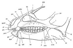
The recent revision of the diagnosis by Norman et al. (2011[5]: 236) included five features. Two of these partially overlap those enumerated in the diagnosis above, namely the absence of enlarged caniniform teeth and reduced ornamentation on cheek tooth crowns. One feature listed by Norman and colleagues (12-14 dentary teeth) is primitive with a broader distribution than Abrictosaurus consors. Another feature (dorsoventrally expanded anterior end of the dentary) is regarded here as a synapomorphy for derived heterodontosaurids including Heterodontosaurus tucki. The absence of a projecting maxillary ridge and buccal emargination, the final feature cited in the revised diagnosis of Norman et al. (2011)[5], is regarded here as an artifact of preservation. The neurovascular openings, everted rim on the maxilla, and form of the opposing depression on the dentary clearly indicate that Abrictosaurus consors had a buccal emargination comparable in depth to that in other heterodontosaurids (Figs 34A, 35). Postmortem transverse compression of the skull has brought the mandibles together and flattened prominent facial features.
Abrictosaurus consors is most similar in cranial and postcranial morphology to Heterodontosaurus tucki. They differ most noticeably in the absence of well-formed caniniform teeth in Abrictosaurus consors and in the shape and ornamentation of noncaniniform teeth. The presence and size of caniniform teeth may be the product of sexual dimorphism (Thulborn 1974[1]), although the rarity of reduced caniniform teeth among the many specimens of heterodontosaurids now in collections argues against this interpretation (Norman et al. 2011[5]).
Description
The holotypic skeleton of Abrictosaurus consors (NHMUK RU B54), which is preserved for the most part in natural articulation, is the most complete specimen of a heterodontosaurid with the single exception of a nearly complete skeleton of Heterodontosaurus tucki (SAM-PK-K1332; Santa Luca et al. 1976[9]; Santa Luca 1980[10]). The following brief description of the holotypic specimen clarifies aspects of the morphology critical to the taxonomy and phylogenetic relationships of this important heterodontosaurid. The skull and skeleton require further preparation before a more detailed description is possible. The skull and femur of the single known specimen of Abrictosaurus consors are approximately 71% and 68% of the length of the skull and femur in Heterodontosaurus tucki (SAM-PK-K1332; Table 3), respectively. This suggests that Abrictosaurus consors probably grew to a comparable adult body size.
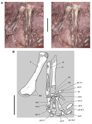
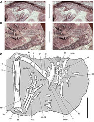
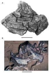
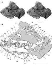
Preserved in two pieces, the skull is transversely compressed and better exposed in left lateral view (Fig. 31). Much of the dorsal skull roof, braincase, and posterior end of the lower jaws are broken away. The postcranial skeleton is preserved on separate blocks, one preserving an articulated right forelimb (Fig. 36) and the other the ilia, an ischium, sacrum and left hindlimb (Fig. 37). The phalanges of the right manus cross onto the skull block and are partially exposed near the scleral ring in the left orbit (Fig. 31).
Premaxilla
Both premaxillae are partially preserved with the right shifted slightly ventral to the left. Portions that are broken on both sides include sections of the alveolar border, the internarial processes, and the distal end of both posterolateral processes (Fig. 31). The posterior end of the alveolar margin on the left side was originally shown as complete (Fig. 34A) but now is damaged (Fig. 31). The premaxillary tooth row is set below the maxillary tooth row, although the “overhanging and hood-like” positioning of the premaxilla (Thulborn 1974[1]: 154; Fig. 34A) appears to have been enhanced by postmortem displacement. The form of the premaxilla is very similar to that in Heterodontosaurus. Although incomplete, the narial fossa is broad and extends toward the alveolar margin (Figs 31, 35).
The posterolateral process of the premaxilla narrows slightly posterior to the external naris and then broadens in transverse width distally, which in Heterodontosaurus is directly related to the anterior margin of the arched premaxilla-maxilla diastema (Fig. 59). In Abrictosaurus the region of a potential arched diastema, however, is incomplete on both sides (Figs 31, 32). The lower dentition would not have necessitated a diastema, as there is no development of the typical heterodontosaurid dentary caniniform tooth (Fig. 35).
Thulborn suggested, nonetheless, that caniniform crowns in heterodontosaurids might be sexually dimorphic, that NHMUK RU B54 may represent a female, and that a deep arched diastema may have been present in Abrictosaurus without an opposing lower caniniform tooth. Norman et al. (2011)[5] presented a similar interpretation (Fig. 34B). Although sexual dimorphism remains a plausible hypothesis, details in the region of the diastema on both sides suggest that only a small diastema could have been present in Abrictosaurus. First, the alveolar margin of the right maxilla, which is preserved farther anteriorly than the left, extends anteriorly as a horizontal border as far as the anterior end of the dentary. There is no hint of an arched embayment on the right side. Second, the left dentary tooth 2, which is positioned below the proposed diastema, is truncated by an oblique wear facet (Fig. 32), indicating tooth-to-tooth contact with an opposing anterior maxillary crown (Figs 31, 32). Although no maxillary crowns are preserved in place above this tooth on either side, this planar wear facet suggests that a maxillary crown dorsal to this tooth was originally present on the anterior maxillary alveolar margin and that it has broken away (Fig. 35). It seems doubtful, finally, that the hypertrophied tooth in a caniniform-diastema complex would be reduced, while the recessed fossa that houses such a crown upon jaw closure would be maintained.
Maxilla, lacrimal, postorbital, and palpebral
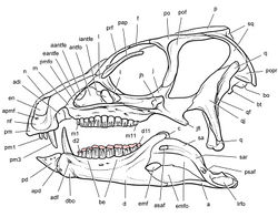
The maxilla has a dorsoventrally deep buccal emargination, which is bordered dorsally by an arched row of large neurovascular foramina and the everted and gently arched external rim of the antorbital fossa. The narrow width of the anterodorsal ramus brings the premaxilla in close proximity to the antorbital fossa as in Heterodontosaurus. At the base of this maxillary ramus, the anterior corner of the external opening of the antorbital fossa has a broader arc than in Heterodontosaurus. The external opening of the antorbital fossa is subtriangular (Fig. 35) as in Heterodontosaurus. As seen in lateral view (Figs 31, 35), the dorsally arched alveolar margin of the maxillary tooth row also resembles the condition in Heterodontosaurus.
The subtriangular lacrimal may be slightly broader dorsally than in Heterodontosaurus, its lateral surface lacking the subtriangular external fossa present in the latter genus. A slender, dorsoventrally flattened palpebral is preserved near its natural articulation with the lacrimal. In cross-section and length, the palpebral closely resembles that in Heterodontosaurus (Figs 31, 35). This bone was previously identified as the prefrontal (Fig. 34).
Quadratojugal and quadrate
The partially preserved quadratojugal and ventral end of the quadrate form a T-shaped junction in Abrictosaurus as in Heterodontosaurus. The jaw joint appears to be preserved in natural articulation (Fig. 31). In Abrictosaurus the jaw articulation is set below the tooth row to the level of a horizontal line passing through the middle of the dentary ramus (Figs 31, 34).
Lower jaw
The dentary has a deep, arched buccal emargination that dissipates anteriorly near the subconical second tooth (Figs 31, 35). An anterior dentary foramen is located farther ventrally under the same tooth as in Heterodontosaurus (Figs 31, 35). The anterior end of the dentary is more strongly expanded dorsoventrally than in Heterodontosaurus, although both are similar in form. The articular surface for the predentary, as in Heterodontosaurus and Pegomastax gen. n. sp. n., is saddle-shaped; in lateral view the articular surface is dorsoventrally convex and transversely concave (Fig. 32). In addition, a rugose swelling here termed a dentary boss is present on the lateral aspect of the anterior end (Figs 32, 59).
The posterior margin of the dentary is not well preserved; the notched posterior margin of the right dentary suggests that an external mandibular fenestra of moderate size may have been present as in Heterodontosaurus tucki (contra Thulborn 1974[1]: 158). An internal mandibular foramen is also present between the splenial and prearticular in medial view of the right mandibular ramus (contra Thulborn 1974[1]: 158; Fig. 31). The splenial tapers anteriorly before reaching the symphysis, where a section of Meckel’s canal is exposed. The canal is represented by a narrow trough running just above the ventral margin of the dentary (Fig. 31) as in Echinodon, Heterodontosaurus and several other heterodontosaurids.
Premaxillary teeth
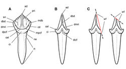
There are three premaxillary crowns as in other heterodontosaurids (rather than two, contra Thulborn 1974[1]: 15) (Fig. 31, 32). The first and smallest of the premaxillary teeth is preserved on the left side, its crown broken away flush with the alveolar margin (Figs 31, 32). This tooth (pm1) is inset a good distance from the anterior end of the premaxilla. Pm2 and 3 are subconical and slightly recurved, their apical ends broken away. Judging from the preserved portions of the crowns, more of the crown tip of pm3 is broken away than in pm2. Their smooth crowns are bounded mesially and distally by low, but distinct, edges. The crown merges with the root without an intervening neck (Fig. 10A), unlike the premaxillary teeth in Echinodon and the first two premaxillary crowns in Heterodontosaurus. In Abrictosaurus and other heterodontosaurids, pm3 is the largest tooth in the premaxillary series, although in Abrictosaurus the size differential is minor. Pm3 is not transversely compressed, and there is no development of serrations on mesial or distal edges of the crown (Fig. 31).
Maxillary teeth
A complete maxillary series probably included 14 teeth, as estimated from the 12 preserved maxillary teeth in the left maxilla and the space between these and the anterior end of the maxilla on the right side (Figs 31, 35). At least two small teeth may have been present at the mesial end of the series, indicating there were more than 12 maxillary teeth (contra Thulborn 1974[1]: 158). The teeth are numbered accordingly, accounting for two missing anterior crowns. In Abrictosaurus, Heterodontosaurus and Echinodon, the tooth rows are nearly straight in apical view, whereas in Lycorhinus and “Geranosaurus” they are bowed medially (Gow 1990[11]; Broom 1911[12]). Opposite to the condition in Echinodon, the maxillary teeth are slightly taller than opposing dentary teeth along the tooth row (Figs 31, 35).
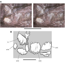
Abrictosaurus has very distinctive maxillary crowns, best exemplified in the middle portion of the tooth rows. The maxillary crowns are extremely tall, their height more than twice their maximum width in the anterior center of the tooth row (teeth 5-10). Crown proportions (height versus width) decrease distally toward the end of the tooth row. Some of the extra crown height of the central teeth is accommodated by an upward arching of the alveolar margin as in Heterodontosaurus, which keeps the axis of occlusion horizontal (Fig. 35). In Echinodon and Lycorhinus, in contrast, the crowns in the tooth row are more similar in size and the alveolar margins less arched.
The cingulum is reduced in Abrictosaurus with the crown in most teeth expanding gently from the root. The best-developed cingulum occurs in smaller teeth at the distal end of maxillary and dentary tooth rows. In these teeth, as in Heterodontosaurus, the crown expands more abruptly from the root (Figs 31, 32).
In labial view, mesial and distal crown edges are nearly parallel, with no development of marginal ridges like those in Echinodon, Lycorhinus, and Heterodontosaurus. The denticulate margin near the top of the crown has approximately five to six denticles to each side. These margins slope at a low angle, about 30 degrees from the perpendicular to the crown axis. The denticulate portion of the crown, thus, is confined to the apical 25% of crown height. In cross-section, the crowns are blade-shaped and transversely narrower than in Lycorhinus and Heterodontosaurus. The enamel may have an asymmetrical distribution, thicker on the labial side of the crown, but this is not well established.
The maxillary crowns are canted at a slight angle to their roots, directing the crown lingually. Low-angle wear facets graze the lingual side of the maxillary crowns. As in Heterodontosaurus more than one facet is present on a single crown, as opposing cheek teeth are not aligned one-to-one. Contrary to Thulborn (1974[1]: 154, 159), the wear facets do not lie in a single plane and two wear facets, rather than only one, occur adjacent to one another on several crowns. Maxillary and dentary teeth did not occlude one-to-one as implied by Thulborn, nor do they occlude in strict alternation as suggested by Weishampel (1984[13]: 53).
There is indisputable evidence for active tooth replacement in Abrictosaurus. The base of the penultimate maxillary tooth on the right side has broken away to expose an erupting crown (Fig. 33). Thulborn (1974[1]: 159) stated there is no evidence of active tooth replacement in Abrictosaurus, suggesting that the entire maxillary and dentary tooth rows were replaced simultaneously during aestivation (Thulborn 1978[14]). The evidence from the dentition does not support this scenario.
Dentary teeth
The left dentary has 14 teeth, which may comprise a complete tooth row (Figs 31, 35). A small distalmost dentary tooth, however, is preserved on the right side that may constitute dentary tooth 15, which could be obscured by matrix on the left side. The first two dentary teeth have crowns with an atypical shape (Figs 32, 35), and these may correspond to the peglike tooth and caniniform tooth in some heterodontosaurids such as Lycorhinus. The small first tooth has a smooth subconical crown that is swollen slightly above the root. The larger second crown, which also shows some swelling above the root, has a convex mesial margin and low unornamented distal carina (Fig. 32). This tooth may represent a reduced caniniform tooth, as it occupies a similar position at the anterior end of the buccal emargination.
The remaining dentary teeth have diamond-shaped crowns that are shorter than opposing maxillary crowns (Fig. 31). The first five dentary tooth crowns do not overlap one another. The remainder of the dentary crowns and all of the maxillary crowns are closely spaced or in contact. As much as 50% of the crown is bordered by mesial and distal denticulate margins, which are more steeply inclined than the denticulate margins of opposing maxillary crowns. Echinodon also exhibits a similar differential in the inclination of the denticulate margins between maxillary and dentary crowns, the latter also more steeply inclined.
Skull reconstruction
The reconstruction of the partial skull and dentition of Abrictosaurus (Fig. 35) differs in several regards from previous reconstructions (Fig. 34; Thulborn 1974[1]; Norman et al. 2011[5]). In the present reconstruction, the premaxilla is rotated in a clockwise direction to restore its position relative to the maxilla and bring the premaxillary tooth row, now with three teeth rather than two, closer to a horizontal orientation. Based on evidence from the right side of the skull, the anterior end of the maxilla is restored, the diastema reduced, and two small teeth added to the anterior end of the tooth row (Fig. 35). The alveolar margins of the dentary and maxillary tooth rows are arched ventrally and dorsally, respectively, as preserved on the skull and as in Heterodontosaurus. Using the better-preserved skulls of Heterodontosaurus as a guide, the palpebral is rotated clockwise to align it with the dorsal margin of the snout. The external opening of the antorbital fenestra is enlarged, following preserved margins on the maxilla and lacrimal. Finally, the ventral margins of the predentary and anterior end of the dentary are reduced and expanded, respectively, as preserved (Fig. 34).
Axial skeleton
The articulated cervical series is only partially exposed. The atlas and axis are incomplete, and C3-9 are exposed in dorsal view. Judging from their neural arches, length decreases markedly from C6-9 as in Heterodontosaurus. The neural arches of C8 and C9 are only approximately one-half that of C3.
In C3 the neural spine is low and ridgelike and the postzygapophyses have smooth dorsal surfaces lacking any development of epipophyseal processes. In Heterodontosaurus, in contrast, a well developed epipophysis projects as a subconical process from the postzygapophysis of C3. The longer, arched postzygapophyses of C5 and shorter postzygapophyses of C6 also lack discrete epipophyseal processes. A low crest at the base of each of these postzygapophyses joins the low neural spine. In C6 the tab-shaped neural spine is located at the anterior end of the neural arch. Although the form of these vertebrae is similar to that in Heterodontosaurus, important differences include the absence of the unusual anterodorsally inclined neural spines in C5 and C6.
Ossified tendons are associated with the neural arches of the dorsal and sacral vertebrae, but as in Heterodontosaurus few of these are in natural position. Ossified tendons appear to be limited to dorsal and sacral regions of the vertebral column, and none is present in the proximal portion of the caudal series (Fig. 37).
Pectoral girdle
The scapula, the only bone of the pectoral girdle that is exposed, shows a prominent acromial expansion and strap-shaped proximal scapular blade. Little else can be said without further preparation.
Forelimb
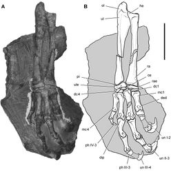
The right forelimb is nearly complete with most of its bones in articulation or near their natural location and with the manus in ventral view overlapping the left orbit of the skull (Fig. 36). The humerus, exposed in anterior view, is poorly preserved proximally and distally. The deltopectoral crest projects strongly from the proximal shaft. The length of the crest is about 34% of humeral length (Table 6). In Heterodontosaurus, the proximal end of the crest arches more abruptly from the shaft than in Abrictosaurus, and the relative length of the crest is greater (approximately 42% of the humerus) (Table 8). The radius and ulna are preserved on both sides but are incomplete distally and only partially exposed (Fig. 36). In Heterodontosaurus the ulnar shaft thickens on its ventral aspect prior to the strong proximally projecting olecranon process. Although that process is not preserved or exposed in Abrictosaurus, the right ulna shows the ventral thickening of the proximal shaft.
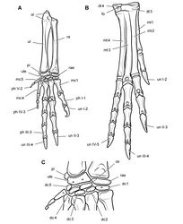
The carpus is composed of many elements, which may have been assembled in articulation with a compact arrangement as in Heterodontosaurus. In the right carpus, three carpals are preserved proximal to metacarpals 3 through 5 (Fig. 36). The large ovoid bone distal to the ulna and closest to the base of metacarpal 5 may represent the ulnare, which in Heterodontosaurus is even larger and more subrectangular in shape. The pair of smaller subspherical elements associated with the bases of metacarpals 3 and 4 may represent distal carpals. In Heterodontosaurus the distal carpals in the lateral side of the carpus appear more lenticular in shape with an overlapping arrangement (Fig. 67C).
The metacarpals are proportionately elongate relative to either the bones of the forearm or humerus as in Heterodontosaurus (Fig. 36). Metacarpal 2, the most complete of the series, is approximately 32% of humeral length (Table 6). The same proportion in Heterodontosaurus is only 27% (Table 8), and thus the hand in Abrictosaurus appears to be proportionately longer. An unusual feature of the metacarpus in Heterodontosaurus is that metacarpal 2 is slightly longer and more robust than metacarpal 3 (Table 8). Their distal condyles, nevertheless, are nearly aligned, as the base of metacarpal 2 is inset slightly into the carpus relative to metacarpal 3 (Fig. 36). The slightly inset position of the base of metacarpal 2 relative to metacarpal 3 also is preserved on both sides in the articulated skeleton of Heterodontosaurus (Fig. 65). Metacarpals 1-3 are more robust and longer than metacarpals 4 and 5, the former measured from the left manus. Metacarpal 5 is the shortest metacarpal with a shorter length than in Heterodontosaurus (Tables 6, 8).
The proximal ends of the metacarpals show edges that appear to square the base of the bone as in Heterodontosaurus and the Kayenta heterodontosaurid. The squared bases articulate against each other in Heterodontosaurus, and a similar condition may hold for at least the medial metacarpals in Abrictosaurus (Fig. 36). In Heterodontosaurus, however, the metacarpus is exposed only in dorsal view (Figs 65, 67). In Abrictosaurus, in contrast, the metacarpus is currently exposed only in ventral view. The distal ends of metacarpals 1-3 are expanded, and thus it seems likely that distal extensor pits would be present dorsally for hyperextension of the phalanges as in Heterodontosaurus.
The right metacarpus, exposed in ventral view, provides the best information regarding the disposition of the digits (Fig. 36). Metacarpal 1 diverges medially only slightly from an axis established on the bones of the forearm. As in Heterodontosaurus (Fig. 67A), the distal condyles are strongly asymmetrical, which cants the phalanges of digit I medially. Digit V, preserved only in the right manus, diverges at nearly a right angle laterally as in Heterodontosaurus and other bipedal ornithischians such as Hypsilophodon (Galton 1974a[15]). The articulation of the base of metacarpal 5 with the lateral aspect of the ulnare, which is also present in Heterodontosaurus, seems to be the natural disposition of this short digit (Figs 36, 67).
The phalangeal formula is poorly known in Abrictosaurus. In the left manus, digit IV has two phalanges, and in the right manus digit V has one (Fig. 36). The distal ends of phalanges IV-2 and V-1 are rounded, and so the presence of a diminutive nonungual terminal phalanx in each digit, as occurs in Heterodontosaurus (Fig. 67),cannot be ruled out. The phalanges of digits I-III are not well exposed. The proximal phalanges of digits I-III show a similar decrease in length to the lateral side as in Heterodontosaurus. The distal ends of phalanges I-1 and II-1 are preserved on the skull block over the left orbit (Fig. 31). It is possible that the remaining phalanges of digits I-III are embedded in matrix within the orbit. The edge of an extremely long recurved element may represent the edge of an ungual. Detailed preparation of the skull block is warranted.
Pelvic girdle
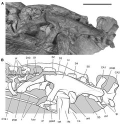
The ilium, preserved in part on both sides, is very similar to that in Heterodontosaurus (Fig. 37, Table 6). The gently everted dorsal margin arches between the slender preacetabular process and the proportionately long postacetabular process. Although incomplete, the anterior end of the preacetabular process appears to taper gradually rather than terminating with a gentle lobe-shaped expansion as in Heterodontosaurus (Santa Luca 1980[10]: fig. 18A). The acetabulum, also similar to that in Heterodontosaurus, is completely open with no development of a supraacetabular rim. The pubic peduncle is longer and narrower than the more robust ischial peduncle, which does not project laterally as prominently as in Heterodontosaurus (Fig. 68). Only the proximal shaft of the right ischium is preserved. No obturator process is visible, but the shaft is not completely exposed (Fig. 37). {| class="wikitable" ; style="width: 100%" |+ Table 6. Measurements (mm) of the holotypic skeleton of the South African heterodontosaurid Abrictosaurus consors (NHMUK RU B54). Measurements are rounded to the nearest millimeter and average right and left sides except where indicated. Parentheses indicate estimated measurement. |- ! Structure !! Measurement !! |- | Skull || Length || (100) |- | Scapula || Blade, minimum width || 5 |- | Humerus || Length || 50 |- | Deltopectoral crest length || 17 |- | Ulna || Length || (39) |- | Radius || Length || (36) |- | Manus || Metacarpal 1 length || 13 |- | Metacarpal 2 length || 16 |- | Metacarpal 3 length || 151 |- | Metacarpal 4 length || (9)1 |- | Metacarpal 5 length || 4 |- | Phalanx I-1 length || 10 |- | Phalanx II-1 length || 8 |- | Phalanx III-1 length || 5 |- | Phalanx V-1 length || 2 |- | Ilium || Blade length || 623 |- | Blade, height dorsal to acetabular rim || 11 |- | Preacetabular process length || 212 |- | Postacetabular process length || 19 |- | Pubic peduncle length || 11 |- | Ischial peduncle length || 7 |- | Femur || Length || 78 |- | Minimum shaft diameter || 8 |- | Head to distal end of fourth trochanter || 30 |- | Tibia || Length || 100 |- | Minimum shaft diameter || 7 |- | Pes || Digit III length || (108) |- | Metatarsal 1 length || 31 |- | Metatarsal 2 length || 47 |- | Metatarsal 3 length || 53 |- | Metatarsal 4 length || (48) |}
Hindlimb
The general form of the femur does not depart significantly from that in Heterodontosaurus (Santa Luca 1980[10], fig. 18B). The shaft is bowed with a pendant fourth trochanter located proximal to mid-shaft (Fig. 37) as in Heterodontosaurus. There is no development of an anterior intercondylar groove. The anterior trochanter, which extends to the level of the greater trochanter, is separated from the shaft of the femur by a deep cleft, as seen in lateral view (Fig. 37). In Heterodontosaurus, in contrast, the lesser trochanter projects dorsally alongside the shaft, to which it is fully coossified (Fig. 68). The greater trochanter in Abrictosaurus is proportionately narrow, subequal in anteroposterior width to the anterior trochanter (Fig. 37). In Heterodontosaurus and neornithischians in general, the greater trochanter is always broader than the lesser trochanter in lateral view. The pendant fourth trochanter is located on the proximal half of the femoral shaft as in Heterodontosaurus.
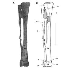
In Heterodontosaurus several features in the crus arose in parallel among coelurosaurian theropods, including an articular crest protruding from the shaft of the proximal end of the tibia to support the reduced shaft of the fibula, reduction of the fibular shaft to a narrow rod, and coossification of the distal ends of the tibia and fibula with the proximal tarsals. Distal coossification of the tibia, fibula, and proximal tarsals (as a “tibiofibulotarsus”), however, may be variable in Heterodontosaurus. A referred right tibia, fibula, and astragalocalcaneum do not exhibit obliterating coossification between the crus and tarsus (Fig. 70). In Abrictosaurus most of the fibular shaft is similarly reduced to a rod, but there does not appear to be any development of a lateral supporting flange on the tibial shaft or distal fusion between the crus and tarsus (Fig. 37).
The tarsus includes the astragalus, calcaneum, and lateral and medial distal tarsals, which do not appear to be coossified (Fig. 37). The flat, platelike medial distal tarsal is preserved in articulation over metatarsals 2 and 3, whereas the smaller cuboid lateral distal tarsal is displaced a short distance from its articulation over metatarsal 4.
The metatarsal shafts are not coossified, and the pes as preserved is less compact than in Heterodontosaurus (Fig. 37). The distal end of metatarsal 3 diverges laterally, and the shaft of metatarsal 4 follows a S-shaped curve. These plesiomorphic features are only weakly expressed in Heterodontosaurus. In Abrictosaurus metatarsal 1 tapers toward its proximal end as an extremely thin splint, which is very narrow both transversely and dorsoventrally. Metatarsal 3 is the longest, with metatarsals 2 and 4 subequal in length. An extensor pit above the distal condyles occurs only on metatarsal 3 rather than on both metatarsals 3 and 4 as in Heterodontosaurus. Extensor pits are also developed above the distal condyles of the proximal phalanges of digits I-III. Only pedal digits I and IV preserve complete phalangeal series, which are consistent with the primitive ornithischian phalangeal formula (2-3-4-5-0).
Taxon Treatment
- Sereno, P; 2012: Taxonomy, morphology, masticatory function and phylogeny of heterodontosaurid dinosaurs ZooKeys, 226: 1-225. doi
Other References
- ↑ 1.00 1.01 1.02 1.03 1.04 1.05 1.06 1.07 1.08 1.09 1.10 1.11 1.12 Thulborn R (1974) A new heterodontosaurid dinosaur (Reptilia: Ornithischia) from the Upper Triassic Red Beds of Lesotho. Zoological Journal of the Linnean Society 55: 151-175. doi: 10.1111/j.1096-3642.1974.tb01591.x
- ↑ 2.0 2.1 Hopson J (1975) On the generic separation of the ornithischian Lycorhinus and Heterodontosaurus from the Stormberg Series (Upper Triassic) of South Africa. South African Journal of Science 71: 302-305.
- ↑ Galton P (1986) Herbivorous adaptations of Late Triassic and Early Jurassic dinosaurs. In: Padian K (Ed). The Beginning of the Age of Dinosaurs. Faunal Change Across the Triassic-Jurassic Boundary. Cambridge University Press, London and New York: 203-221.
- ↑ Smith J (1997) Heterodontosauridae. In: Currie P Padian K (Eds). Encyclopedia of Dinosaurs. Academic Press, San Diego: 317-320.
- ↑ 5.0 5.1 5.2 5.3 5.4 5.5 5.6 Norman D, Crompton A, Butler R, Porro L, Charig A (2011) The Lower Jurassic ornithischian dinosaur Heterodontosaurus tucki Crompton and Charig 1962: cranial anatomy, functional morphology, taxonomy, and relationships. Zoological Journal of the Linnean Society 162: 182-279.
- ↑ Kitching J, Raath M (1984) Fossils from the Elliot and Clarens Formations (Karoo Sequence) of the northeastern Cape, Orange Free State and Lesotho, and a suggested biozonation based on tetrapods. Palaeontologia africana 25: 111-125.
- ↑ Knoll F (2005) The tetrapod fauna of the Upper Elliot and Clarens formations in the main Karoo Basin (South Africa and Lesotho). Bulletin de la Société géologique de France 176: 81-91. doi: 10.2113/176.1.81
- ↑ Gradstein F, Ogg J (2009) The geologic time scale. In: Hedges S Kumar S (Eds). , The Timetree of Life. Oxford University Press, Oxford: 26-34.
- ↑ Santa Luca A, Crompton A, Charig A (1976) A complete skeleton of the Late Triassic ornithischian Heterodontosaurus tucki. Nature 264: 324-328.
- ↑ 10.0 10.1 10.2 Santa Luca A (1980) The postcranial skeleton of Heterodontosaurus tucki (Reptilia, Ornithischia) from the Stormberg of South Africa. Annals of the South African Museum 79: 159–211. doi: 10.1038/264324a0
- ↑ Gow C (1990) A tooth-bearing maxilla referable to Lycorhinus angustidens Haughton, 1924 (Dinosauria, Ornithischia). Annals of the South African Museum 99: 367-380.
- ↑ Broom R (1911) On the dinosaurs of the Stormberg, South Africa. Annals of the South African Museum 7: 291-307.
- ↑ Weishampel D (1984) Evolution of jaw mechanisms in ornithopod dinosaurs. Advances in Anatomy and Cell Biology 87: 1-109. doi: 10.1007/978-3-642-69533-9_1
- ↑ Thulborn R (1978) Aestivation among ornithopod dinosaurs of the African Trias. Lethaia 11: 185-198. doi: 10.1111/j.1502-3931.1978.tb01226.x
- ↑ Galton P (1974a) The ornithischian dinosaur Hypsilophodon from the Wealden of the Isle of Wight. Bulletin, British Museum (Natural History) Geology 25: 1-152.
Images
|
- ↑ Thulborn R (1974) A new heterodontosaurid dinosaur (Reptilia: Ornithischia) from the Upper Triassic Red Beds of Lesotho. Zoological Journal of the Linnean Society 55: 151-175. doi: 10.1111/j.1096-3642.1974.tb01591.x
- ↑ Norman D, Crompton A, Butler R, Porro L, Charig A (2011) The Lower Jurassic ornithischian dinosaur Heterodontosaurus tucki Crompton and Charig 1962: cranial anatomy, functional morphology, taxonomy, and relationships. Zoological Journal of the Linnean Society 162: 182-279.

![Figure 34. Skull of Abrictosaurus consors from the Lower Jurassic Upper Elliot Formation of South Africa. A Skull bones of NHMUK RU B54 in left lateral view as initially identified (from Thulborn 1974[1]). B Diagrammatic skull reconstruction in left lateral view based on NHMUK RU B54 (from Norman et al. 2011[2]). Scale bar equals 1 cm in A. Abbreviations: a angular adi arched diastema antfo antorbital fossa d dentary en external nares j jugal l lacrimal or left m maxilla or orbit pap palpebral pd predentary pf prefrontal pm premaxilla q quadrate qj quadratojugal r right sa surangular scr scleral ring.](https://species-id.net/o/thumb.php?f=ZooKeys-226-001-g034.jpg&width=209)
