Difference between revisions of "Ectinorus spiculatus"
m (Imported from ZooKeys) |
m (1 revision) |
(No difference)
| |
Latest revision as of 15:25, 18 August 2011
| Notice: | This page is derived from the original publication listed below, whose author(s) should always be credited. Further contributors may edit and improve the content of this page and, consequently, need to be credited as well (see page history). Any assessment of factual correctness requires a careful review of the original article as well as of subsequent contributions.
If you are uncertain whether your planned contribution is correct or not, we suggest that you use the associated discussion page instead of editing the page directly. This page should be cited as follows (rationale):
Citation formats to copy and paste
BibTeX: @article{Hastriter2011ZooKeys124, RIS/ Endnote: TY - JOUR Wikipedia/ Citizendium: <ref name="Hastriter2011ZooKeys124">{{Citation See also the citation download page at the journal. |
Ordo: Siphonaptera
Familia: Rhopalopsyllidae
Genus: Ectinorus
Name
Ectinorus spiculatus Hastriter and Sage sp. n. – Wikispecies link – ZooBank link – Pensoft Profile
Type Material
Argentina, Neuquén Province:, 1 km SSW from Route 40 on dirt road to Estancia Llamuco (38°44'1.2"S, 70°17'55.26"W ), vegetation on sandy soil with basaltic rimrock, 1074m, ex Phyllotis xanthopygus (♀), 14 IV 2008, R.D. Sage, Holotype ♂ (RDS-18861); Laguna Blanca National Park, Locality 76, 2.24 km W, 3.12 km S Cerro Mellizo Sud, (39°6' 10.92"S, 70°19'33.42"W ), lava outcrops with Colliguaja sp., 1320m, ex Phyllotis xanthopygus (♂), 14 III 2007, R.D. Sage, allotype ♀ (RDS-18407); same data as allotype except ex Akodon iniscatus (♂), 13 III 2007, paratype ♀ (RDS-18403). Holotype and allotype are deposited in the Museo de Ciencias Naturales “Bernardino Rivadavia" de la Ciudad de Buenos Aires, Republica Argentina; paratype ♀ deposited in the Monte L. Bean Life Science Museum, Brigham Young University, Provo, Utah, U.S.A.
Diagnosis
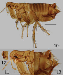
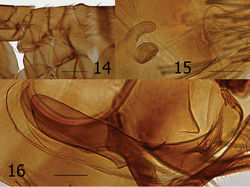
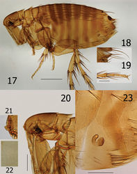
Males key to Ectinorus hertigi (Johnson) in Smit’s (1987:78) key, while females key to Ectinorus barrerai Jordan. Morphologically the male is closely allied with Ectinorus hertigi but may be distinguished from it and all other species of the subgenus Ectinorus by the bilobed apex of the basimere and details of the aedeagus (Figs 13, 16). The presence of seven segments in the labial palpus of females (male with five) is the basis for its similarity with Ectinorus barrerai; however, their similarity is limited. If one continues onward in the key using five segments in the labial palpus (versus 6 to 8), females key out to Ectinorus hertigi also. Females share many similarities with Ectinorus hertigi (few with Ectinorus barrerai) for which they may be separated by an oblique flattened region of the spermatheca at the cribriform area and a very long bursa copulatrix that is reflected postad in a semi-circular arc (Fig. 23).
Description
Chaetotaxy and structural references include only one side of specimen. Head (Figs 10–12). Frons evenly rounded; thickened throughout. Frontal tubercle quadrate; capsule heavily sclerotized but thin caudad. Two placoids between frontal tubercle and sclerotized antennal suture. Eye large, darkly pigmented, sinuate. Ocular setae four; laterals large, middle two much smaller. Tentorium clearly visible anterior to eye. Preantennal setae; one near oral angle, two (large and small) anterior to eye. Third segment of maxillary palpus shorter than others; maxilla acutely sharp. Five segmented labial palpus extending to apex of coxa, apical two segments twice length of either second or third segments; apex blunt with array of fine setae. Antennal scape with apical row of six fine setae; pedicel with three minute dorsal setae; clavus extending onto prosternasome. Post-antennal area with four rows of setae (1, 1, 1, 6 plus intercalaries; female with only two minute setae anterior to main row). Two placoids; occipital groove moderately deep. Row of 18 setules along dorsal margin of antennal groove. Genal lobe bluntly rounded with three small apical setae; five larger marginal setae below eye. Thorax (Figs 10, 11, 14). Pro-, meso-, and metanota each with two rows setae. Eleven to 12 pseudosetae under mesonotal colar. Dorsal apex of metanotum curled downward. Cervical link-plate truncate at apex. Prosternasome grooved for retention of antennal apex; without setae. Mesepimeron with four setae and mesepisternum with two; mesosternum heavily sclerotized along ventral margin with incomplete suture between mesepisternum. Pleural rod bifurcate dorsally. Lateral metanotal area with two large, two small setae. Pleural arch and ridge well developed. Metepisternum and metasternum, fused into one; one large seta. Furca long and delicate. Metepimeron with two vertical rows of setae; anterior with two (dorsal minute), posterior of three (same arrangement in female). Legs (Figs 10, 19). Fore coxa with 28 lateral setae; one long seta at posterior margin. Oblique break mid coxa indicated only at ventro-caudal margin. Two guard setae at femoral-tibial joints; lateral of two long equal on fore femur; shorter on mid and hind femora. Fore and mid femora with two lateral rows of setae; hind femur with single lateral row of 12 setae. Lateral sculpturing of hind femur very fine. Margin of fore, mid, and hind tibiae with 5, 6, and 6 dorsal notches, respectively. Number of setae in respective dorsal notches: fore tibia (beginning with proximal notch) (2, 2, 3, 2, 3), mid tibia (2, 2, 2, 3, 2, 3), hind tibia (2, 2, 2, 2, 2, 3). Lateral setae of each tibia, respectively (6, 6, 8). Inner (mesal) surface of hind tibia adorned with spicules. First hind tarsus with three long setae; two extending to and one extending beyond segment three. Second hind tarsus with two setae extending beyond distotarsomere. Distotarsomeres with four pair lateral plantar bristles; apical pair smallest. Pre-apical plantar bristles two; one small, one larger. Ungue symmetrical. Unmodified Abdominal Segments (Fig. 10). Dorsum of tergum I heavily sclerotized with distinct hump (absent in female); anterior lateral margin thick and sclerotized. Two rows setae. Terga II–III with two rows setae; terga IV–VII with single row. Ventral most setae of terga II–VII not extending below level of round spiracles. Single antesensilial bristles extending from pedestal beneath apical flange of tergum VII. Sternum II with lateral patch of 7–8 small setae. Sterna II-VII with single rows of setae (1, 2, 3, 3, 3, 3). Modified Abdominal Segments (Male) (Figs 10, 13). Sensilium with 17 sensilial pits; surrounded by wide sclerotized area bearing single seta on caudal margin. Spiracle VIII vermiform, curved upward with three small setae dorsad. Tergum VIII large and highly specialized; lateral and apical surfaces with coarsely reticulated pattern. Tergum VIII envelops basal portion of basimere while curling under and behind apical portion of basimere and telomere to form an unusual conical sharp lobe. Caudal margin is adorned with eleven long setae; ventro-caudal margin with two long setae and smaller marginal setae cephalad. Sternum VIII with lateral row of eight long setae; ventral apex with thick incrassation. Dorsad to incrassation extends a moderately sharp projection. Apex of basimere (tergum IX) with two asymmetrical lobes divided by a deep sinus. Dorsal lobe of basimere with numerous setae; ventral lobe with two stout setae. Robust processus basimeris ventralis present; group of stout setae at apex. Length of telomere more than five times width; bluntly rounded at apex, sides parallel. Numerous small setae line margin. Manubrium tapered, curving upward to acute point. Lateral portion of basimere with triangular, darkly sclerotized, caudally directed structure (Fig. 16). (A patch of fine setae are present on each side and appear to be present on a lobe ventrally located on the ventral margin of tergum VIII and may be associated with triangular sclerotization above. (Without dissection of genitalia, this anatomy could not be deciphered for certain). Distal arm of sternum IX long with parallel sides, expanding at tip; lateral setae present on upper third. Notable group of 9–10 long setae on caudally expanded lobe. Vestigial tendon of sternum IX affixed to apical sclerotization of sternum VIII. Aedeagus (Figs 10, 13, 16). Similar to that of Ectinorus hertigi, but median dorsal lobe greatly reduced and lateral lobes expanded. Dorsal armature immense (seen behind basal portion of telomere sandwiched between conical lobe of tergum VIII, Fig. 13), ventral armature reduced. Sclerotized inner tube long, slightly curved ventrad; with annular ring at midpoint. Aedeagal apodeme bluntly rounded at apex; penis rods barely extend beyond apex of apodeme. Modified Abdominal Segments (Female) (Figs 15, 17, 18, 23). Seventh sternum with lateral row of five setae; caudal margin entire with ventral margin incised, creating an indistinct rounded ventral lobe. Single antesensilial bristle arising from strong pedicel. Tergum VIII with group of eight setae above spiracle VIII. Spiracle VIII vermiform, slightly ballooned at base. Lateral row of six long setae on tergum VIII; marginal group of 20 plus setae at apical margin. Sternum VIII with apical rounded lobe, without setae. Sensilium with broad sclerotized ring; 16 sensilial pits. Anal stylet with apical long seta plus one seta longer than anal stylet. Length of anal stylet twice width. Hilla twice length of bulga; hilla approximate width of bulga. Bulga flattened on cribriform region; cribriform area not protruding into bulga. Bursa copulatrix extremely long; curved caudally in circular arc.
Length: Male holotype: 2185µ; female allotype: 2533µ; and female paratype: 2175µ.
Etymology
The specific epithet spiculatus is derived from the characteristic presence of spicules on the mesal surface of the hind tibia.
Remarks
The single male and two females were all collected from different host specimens. The authors feel confident that both sexes belong to the same taxon for the following reasons: 1) Both male and female have spicules on the mesal surface of the hind tibiae, 2) both sexes have very similar head chaetotaxy and shape of the genal lobe, 3) the second tarsal segment possesses three long setae, two of which extend beyond segment four, 4) a pair was collected at the same locality (Laguna Blanca National Park) and the other female within close proximity, within 35 km, 5) the male at one locality and female from the other were from the same host species (Phyllotis xanthopygus), and 6) terraine, habitat, and elevations for both localities were nearly the same.
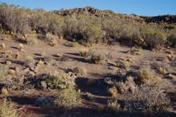
The holotype (RDS-18861) was collected from Phyllotis xanthopygus along the edge of a rimrock of a dark-red basaltic flow from a nearby, unnamed cinder cone (Fig. 26). Deep drifts of unconsolidated, wind-blown sand, filled the fissures in this broken-rock habitat. A dense growth of Colliguaja integerrima Gillies & Hook (“coliguay”) and bunch grasses were the dominant plants. The area was cold, dry, and at times very windy. Only Phyllotis xanthopygus and an undescribed species of Ctenomys (“tuco-tuco”) were trapped at the type locality. It should be noted that Ectinorus lareschaei Hastriter and Sage, 2009 was also collected from the same host specimen as this holotype. Paratypes RDS-18403 and RDS-18407, were collected from Akodon iniscatus and Phyllotis xanthopygus, respectively, in Laguna Blanca National Park at the southern edge of the lava flow forming the cinder cone volcano, Cerro Mellizo Sud. Deep sandy soil fills in the small fissures in the lava flow and there is a sparse growth of the Patagonian steppe vegetation, mostly bunch grasses and smaller shrubs such as Ctenomys integerrima and Nassauvia glomerulosa. Phyllotis xanthopygus was the more common of the five small mammals trapped here, including the mouse opossum Thylamys pallidior (Thomas).
Original Description
- Hastriter, M; Sage, R; 2011: Description of a new species of Ectinorus (E. spiculatus) (Siphonaptera, Rhopalopsyllidae) from Argentina and a review of the subgenus Ichyonus Smit, 1987 ZooKeys, 124: 1-18. doi
Images
|


